File:Cullen1916 fig04.jpg

Original file (1,280 × 1,257 pixels, file size: 508 KB, MIME type: image/jpeg)
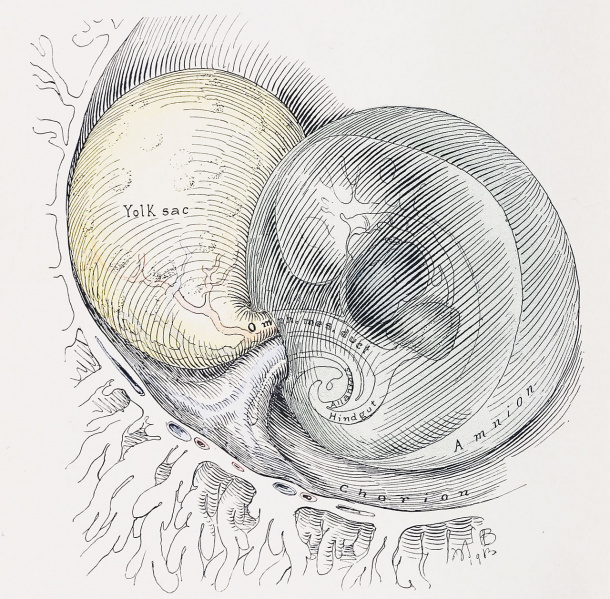
Fig. 4. A Diagrammatic Representation of a Human Embryo, about 3.5 mm. Long
Showing the Effect of the Expanding Amnion upon the Yolk-sac and Body-stalk.
The amnion has now completely encircled the embryo, and with its increase in size has crowded the yolk-sac away. Outside the amnion the yolk-sac and body-stalk are separate structures; inside the amniotic ring they are fused, forming the umbilical cord. The amnion covers the cord as far as the embryo. It is probable that the tension produced on the yolk-Stalk by the amnion contributes to the primitive kink in the alimentary canal.
Reference
Cullen TS. Embryology, anatomy, and diseases of the umbilicus together with diseases of the urachus. (1916) W. B. Saunders Company, Philadelphia And London.
Cite this page: Hill, M.A. (2024, May 18) Embryology Cullen1916 fig04.jpg. Retrieved from https://embryology.med.unsw.edu.au/embryology/index.php/File:Cullen1916_fig04.jpg
- © Dr Mark Hill 2024, UNSW Embryology ISBN: 978 0 7334 2609 4 - UNSW CRICOS Provider Code No. 00098G
File history
Click on a date/time to view the file as it appeared at that time.
| Date/Time | Thumbnail | Dimensions | User | Comment | |
|---|---|---|---|---|---|
| current | 16:25, 27 October 2018 |  | 1,280 × 1,257 (508 KB) | Z8600021 (talk | contribs) | |
| 16:23, 27 October 2018 |  | 2,054 × 2,180 (1,009 KB) | Z8600021 (talk | contribs) |
You cannot overwrite this file.
File usage
The following 3 pages use this file: