File:Cullen1916 fig03.jpg

Original file (1,280 × 1,443 pixels, file size: 494 KB, MIME type: image/jpeg)
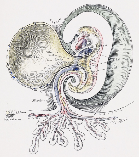
Fig. 3. A Composite Picture Showing the Formation of the Umbilicus in an Embryo 2.5 mm. Long
This drawing was made after a careful study of several embryos of this length described in the literature. Each author was interested in some particular region, but not in the embryo as a whole. The yolk-sac has now become greatly narrowed at its entrance into the body. The narrowed portion is now referred to as the vitelline or omphalomesenteric duct. In its wall are seen the right omphalomesenteric vessels. The digestive tract already shows welldefined differentiation. The allantois now opens into the cloaca. The amnion almost completely encircles the embryo, and in so doing has combined the vitelline duct 'with the body-stalk, containing the chorionic vessels and the allantois, into a common cord. As the development advances this cord will become more compact, thinner, and longer. The exocoelom has been drawn into the embryo and will later unite with the coelom of the pleuroperitoneal cavity.
Reference
Cullen TS. Embryology, anatomy, and diseases of the umbilicus together with diseases of the urachus. (1916) W. B. Saunders Company, Philadelphia And London.
Cite this page: Hill, M.A. (2024, May 19) Embryology Cullen1916 fig03.jpg. Retrieved from https://embryology.med.unsw.edu.au/embryology/index.php/File:Cullen1916_fig03.jpg
- © Dr Mark Hill 2024, UNSW Embryology ISBN: 978 0 7334 2609 4 - UNSW CRICOS Provider Code No. 00098G
File history
Click on a date/time to view the file as it appeared at that time.
| Date/Time | Thumbnail | Dimensions | User | Comment | |
|---|---|---|---|---|---|
| current | 16:20, 27 October 2018 |  | 1,280 × 1,443 (494 KB) | Z8600021 (talk | contribs) | |
| 16:17, 27 October 2018 |  | 2,094 × 2,266 (924 KB) | Z8600021 (talk | contribs) |
You cannot overwrite this file.
File usage
The following 3 pages use this file: