File:Crowder1957 plate01.jpg

Original file (1,280 × 1,457 pixels, file size: 548 KB, MIME type: image/jpeg)
Plate 1
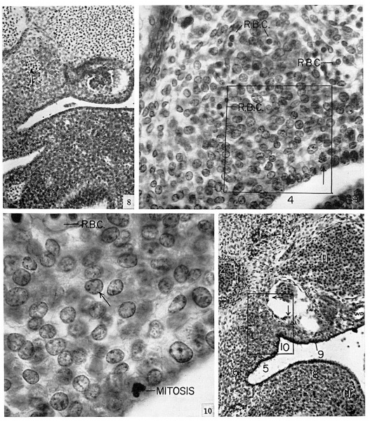
Fig. 8. Transverse section of embryo from beginning of 5th week: midgastric level. Aorta with origin of a mesonephric artery, upper left. Postcardinal vein, upper right. Beneath it, mesonephros with genital ridge on its peritoneal surface. Arrow indicates a transient vessel. Beneath it and mesonephros, enlarged cells define adrenal primordium. The adjacent celomic epithelium can be distinguished from other mesenchyme. No. 3385, 14-3-1. Horizon xv. x150. 8.3 mm.
Fig. 9. From a section taken ahout 150 microns caudal to that of figure 8. Primordiunl diFlerentiated from surrounding mesencliyme by its crowded type C-l cells (see p. I97) with large nuclei and more abundant cytoplasm. Arrow indicates a mitosis. R. 1)’. C., red hlood corpuscles marking capillary plexus on medial surface of primordium. No. 3385, is-I-5. x500.
Fig. 10. Area enclosed by rectangle in figure 9. All cells of type C-1. Cytoplasm abundant, and nuclei large with relatively little chromatin and an eccentric nucleolus. Arrow indicates nucleus showing highly refractile spheroid hotly (see Hg. 38, pl. 8). Zeiss 3.0 mm. apochromat, ap. I40. x1000.
Fig. 11. From a more advanced embryo, showing primordiuni of adrenal protruding into celom. l\-lesonepliros with Malpighian bodies. tubules, and Woffiian duct (IV. 1).). Increase in size of ganglion of sympathetic chain (I3) due to cell division and in- growth of nerve fibers. Two groups of cells in l5owtnan's capsule indicated hy arrows mark a significant differentiation (see fig. .2: fig. 12, pl. 2). No. 721, I4-3-5. Horizon xv. X150. 4. aorta: 5. celomic cavity; 6, mesogastrium: H. stomach; 9, genital ridge; to. adrenal groove: II, postcardinal vein.
- Links: fig 1 | fig 2 | fig 3 | fig 4 | fig 5 | fig 6 | fig 7 | fig 1-7 | plate 1 | plate 2 | plate 3 | 1957 Crowder | Adrenal Development
References
Crowder RE. The development of the adrenal gland in man, with special reference to origin and ultimate location of cell types and evidence in favor of the "cell migration" theory. (1957) Contrib. Embryol., Carnegie Inst. Wash. 36, 193-210.
Cite this page: Hill, M.A. (2024, April 27) Embryology Crowder1957 plate01.jpg. Retrieved from https://embryology.med.unsw.edu.au/embryology/index.php/File:Crowder1957_plate01.jpg
- © Dr Mark Hill 2024, UNSW Embryology ISBN: 978 0 7334 2609 4 - UNSW CRICOS Provider Code No. 00098G
File history
Click on a date/time to view the file as it appeared at that time.
| Date/Time | Thumbnail | Dimensions | User | Comment | |
|---|---|---|---|---|---|
| current | 21:01, 21 June 2017 |  | 1,280 × 1,457 (548 KB) | Z8600021 (talk | contribs) | |
| 21:00, 21 June 2017 |  | 2,161 × 2,984 (1.45 MB) | Z8600021 (talk | contribs) | {{Ref-Crowder1957}} |
You cannot overwrite this file.
File usage
The following page uses this file: