File:Crowder1957 fig01-07.jpg: Difference between revisions
({{Crowder1957 figures}}) |
mNo edit summary |
||
| (One intermediate revision by the same user not shown) | |||
| Line 1: | Line 1: | ||
==Fig. 1 -7== | |||
Figures 1 to 5, drawn to the same scale, X too, illustrate the relative size of the adrenal in horizons xiv to xvii, compared with surrounding structures. Figures 6 and 7 represent the disruption of the cortex by the neural elements in horizon xxiii. | |||
<gallery> | |||
File:Crowder1957 fig01.jpg|Fig. 1 | |||
File:Crowder1957 fig02.jpg|Fig. 2 | |||
File:Crowder1957 fig03.jpg|Fig. 3 | |||
File:Crowder1957 fig04.jpg|Fig. 4 | |||
File:Crowder1957 fig05.jpg|Fig. 5 | |||
File:Crowder1957 fig06.jpg|Fig. 6 | |||
File:Crowder1957 fig07.jpg|Fig. 7 | |||
</gallery> | |||
Fig. 1. Earliest evidence of adrenal primordium and change in character of cells in celomic epithelium over adrenal and g0n:I(_lal areas. | |||
Fig. 2. Genital and adrenal ridges are formed, with adrenal groove separating them. The two groups of cells in the medial wall of Bowman's capsule have tlilierentiated preliminary to migration shown in figure 3. Active proliferation in the sympathetic ganglion is beginning. | |||
Fig. 3. Active migration of cells from I:'»owman's capsule. Beginning invasion of primordium by sympathetic neural elements. | |||
Fig. 4. Massive growth of sympathetic elements. So-called “permanent" cortex. Cell types C-I, C-ll, C-III are present in it. | |||
Fig. 5. Height of sympathetic invasion. Cortex pattern is disrupted by sympathetic elements. Rim of cortex is still intact along the celomic border. Cells from Bowman's capsule and celomic epithelium are still being added to the primordium. Islands of cortical cells are scattered among the neural elements. | |||
Fig. 6. Conditions near the caudal pole of the adrenal at the beginning of the second impetus to growth of sympathetic nerve elements. Much of the cortex is displaced. No capsule is yet formed in this area. | |||
Fig. 7. Condition of the gland when proliferation of sympathetic neural elements is most active. The cortical cords are pushed aside to make room for the nerves and chromaflin elements. The retiform pattern of the sympathetic is well illus. trated. I’ o 1%} .2 '3; | |||
{{Crowder1957 figures}} | {{Crowder1957 figures}} | ||
Latest revision as of 22:31, 21 June 2017
Fig. 1 -7
Figures 1 to 5, drawn to the same scale, X too, illustrate the relative size of the adrenal in horizons xiv to xvii, compared with surrounding structures. Figures 6 and 7 represent the disruption of the cortex by the neural elements in horizon xxiii.
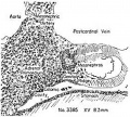
Fig. 1
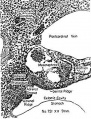
Fig. 2
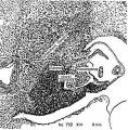
Fig. 3
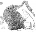
Fig. 4
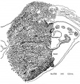
Fig. 5
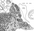
Fig. 6
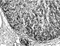
Fig. 7


Fig. 1. Earliest evidence of adrenal primordium and change in character of cells in celomic epithelium over adrenal and g0n:I(_lal areas.
Fig. 2. Genital and adrenal ridges are formed, with adrenal groove separating them. The two groups of cells in the medial wall of Bowman's capsule have tlilierentiated preliminary to migration shown in figure 3. Active proliferation in the sympathetic ganglion is beginning.
Fig. 3. Active migration of cells from I:'»owman's capsule. Beginning invasion of primordium by sympathetic neural elements.
Fig. 4. Massive growth of sympathetic elements. So-called “permanent" cortex. Cell types C-I, C-ll, C-III are present in it.
Fig. 5. Height of sympathetic invasion. Cortex pattern is disrupted by sympathetic elements. Rim of cortex is still intact along the celomic border. Cells from Bowman's capsule and celomic epithelium are still being added to the primordium. Islands of cortical cells are scattered among the neural elements.
Fig. 6. Conditions near the caudal pole of the adrenal at the beginning of the second impetus to growth of sympathetic nerve elements. Much of the cortex is displaced. No capsule is yet formed in this area.
Fig. 7. Condition of the gland when proliferation of sympathetic neural elements is most active. The cortical cords are pushed aside to make room for the nerves and chromaflin elements. The retiform pattern of the sympathetic is well illus. trated. I’ o 1%} .2 '3;
- Links: fig 1 | fig 2 | fig 3 | fig 4 | fig 5 | fig 6 | fig 7 | fig 1-7 | plate 1 | plate 2 | plate 3 | 1957 Crowder | Adrenal Development
References
Crowder RE. The development of the adrenal gland in man, with special reference to origin and ultimate location of cell types and evidence in favor of the "cell migration" theory. (1957) Contrib. Embryol., Carnegie Inst. Wash. 36, 193-210.
Cite this page: Hill, M.A. (2024, May 17) Embryology Crowder1957 fig01-07.jpg. Retrieved from https://embryology.med.unsw.edu.au/embryology/index.php/File:Crowder1957_fig01-07.jpg
- © Dr Mark Hill 2024, UNSW Embryology ISBN: 978 0 7334 2609 4 - UNSW CRICOS Provider Code No. 00098G
File history
Click on a date/time to view the file as it appeared at that time.
| Date/Time | Thumbnail | Dimensions | User | Comment | |
|---|---|---|---|---|---|
| current | 22:03, 21 June 2017 | 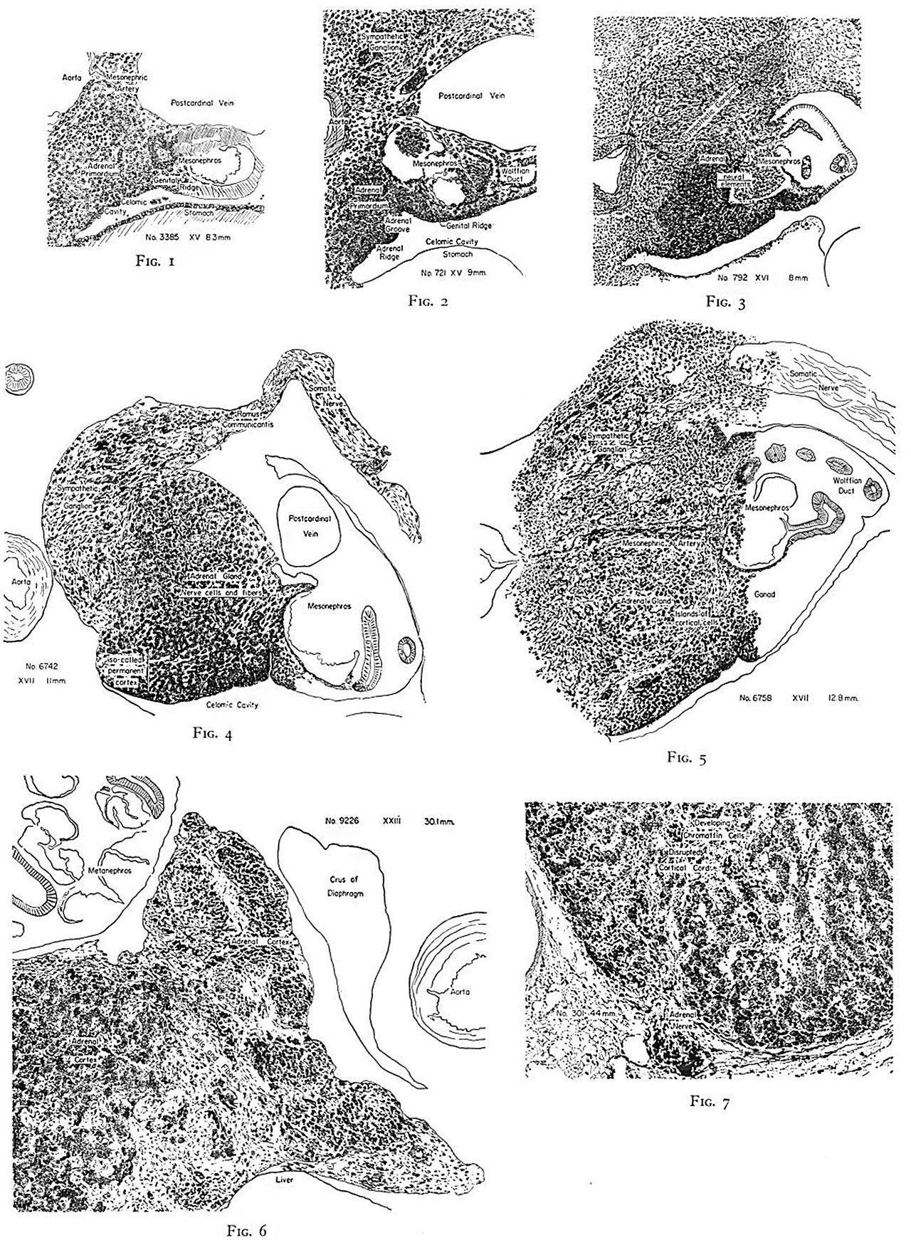 | 1,280 × 1,742 (689 KB) | Z8600021 (talk | contribs) | {{Crowder1957 figures}} |
You cannot overwrite this file.
File usage
The following page uses this file: