File:Cranial neural crest skeletal fate 01.jpg

Original file (800 × 633 pixels, file size: 59 KB, MIME type: image/jpeg)
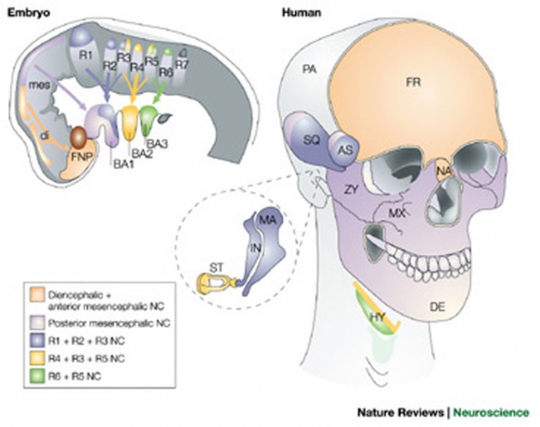
Skeletal fate of cranial neural crest cells in vertebrates
The embryo figure shows colonization of the head and pharyngeal arches by diencephalic, anterior and posterior mesencephalic, and rhombencephalic neural crest cells (NCCs), as indicated by the colour code. The diagram is representative human embryos, although the NCC migratory pathways might differ slightly in different species. The skull drawings show contributions of NCC populations to cranial skeletal elements of humans, based on NCC fate-mapping studies and on extrapolation of avian and mouse data to known homologues in the human.
Some bones, including the squamosal (SQ), alisphenoid (AS), and pterygoid (PT), are shown with mixed contribution from different NCC populations. Note that in mammals the frontal (FR) and parietal (PA) bones have been reported to be of neural crest and mesodermal origin, respectively.
In birds, the frontal and parietal bones have been reported to be either entirely derived from NCCs, or derived from a dual neural crest/mesodermal origin.
Legend
AN, angular bone; AR, articular bone; BA, basihyal; BA1–BA3, pharyngeal arches 1–3; CB, ceratobranchial; CO, columella; DE, dentary bone; di, diencephalon; EB, epibranchial; EN, entoglossum; FNP, frontonasal process; HY, hyoid bone; IN, incus; IS, interorbital septum; JU, jugal bone; MA, malleus; mes, mesencephalon; MX, maxillary bone; NA, nasal bone; NC, nasal capsule; PL, palatine bone; PM, premaxillary bone; QU, quadrate; RP, retroarticular process; R1–R7, rhombomeres 1–7; SO, scleral ossicles; ST, stapes; ZY, zygomatic bone.
(text modified from original figure legend)
FIGURE 1 Nrn1221-f1.jpg
Reference
File history
Click on a date/time to view the file as it appeared at that time.
| Date/Time | Thumbnail | Dimensions | User | Comment | |
|---|---|---|---|---|---|
| current | 11:07, 16 May 2011 |  | 800 × 633 (59 KB) | S8600021 (talk | contribs) | ==Skeletal fate of cranial neural crest cells in vertebrates== The embryo figure shows colonization of the head and pharyngeal arches by diencephalic, anterior and posterior mesencephalic, and rhombencephalic neural crest cells (NCCs), as indicated by th |
You cannot overwrite this file.
File usage
The following 5 pages use this file: