File:Corneal epithelial cells 01.jpg
Corneal_epithelial_cells_01.jpg (800 × 540 pixels, file size: 49 KB, MIME type: image/jpeg)
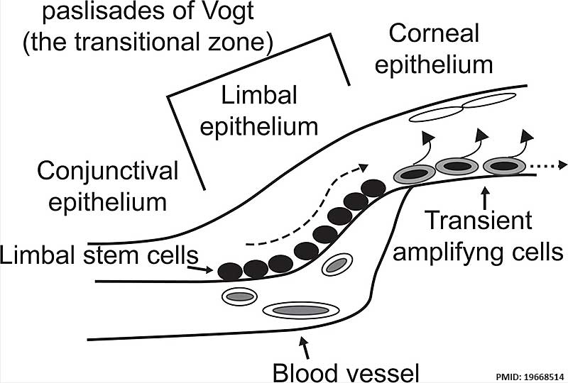
Corneal Epithelial Cells cartoon
The ocular surface is composed of three epithelia - conjunctival, limbal and corneal.
- Limbal stem cells are located in the palisades of Vogt, the transitional zone between the cornea and the conjunctiva.
- Limbal stem cells are close to blood vessels.
- They generate transient amplifying cells that terminally differentiate after a discrete number of cell divisions to corneal epithelial cells and undergo both centripetal migration and vertical migration.
- Links: Cornea Development
Reference
<pubmed>19668514</pubmed>
Copyright
Dovepress Open Access - Articles published under this arrangement are made freely available online upon publication without subscription barriers to access. Users of such published articles are entitled to use, reproduce, disseminate, or display these articles provided that: The original authorship is properly and fully attributed; The journal and publisher are attributed as the original place of publication with correct citation details given; If an original work is subsequently reproduced or disseminated not in its entirety but only in part or as a derivative work this is clearly indicated; No articles are reproduced for commercial use without the prior consent of DMP and payment to DMP of any appropriate fee. Rights of Authors, Readers and the Publisher
File history
Click on a date/time to view the file as it appeared at that time.
| Date/Time | Thumbnail | Dimensions | User | Comment | |
|---|---|---|---|---|---|
| current | 11:40, 30 August 2014 | | 800 × 540 (49 KB) | Z8600021 (talk | contribs) | Corneal Epithelial Cells cartoon== The ocular surface is composed of three epithela, conjunctival, limbal and corneal. Limbal stem cells are located in the palisades of Vogt, the transitional zone between the cornea and the conjunctiva. Limbal stem ce... |
You cannot overwrite this file.
File usage
The following page uses this file: