File:Cooper1938 fig04.jpg: Difference between revisions
From Embryology
(==Plate XXVI== Fig. 4. Single erythrocyte (A) surrounded by mesodermic cells. 60 mm embryo. X 700. Fig. 5. On right is a “primaries ” of the first generation, with cartilage. On the left are “ primaries ” of the second generation. 68 mm embry...) |
mNo edit summary |
||
| Line 1: | Line 1: | ||
== | ==Fig. 4. Single erythrocyte (A) surrounded by mesodermic cells 60 mm embryo== | ||
X 700. | |||
{{Historic Disclaimer}} | {{Historic Disclaimer}} | ||
Latest revision as of 12:54, 27 November 2016
Fig. 4. Single erythrocyte (A) surrounded by mesodermic cells 60 mm embryo
X 700.
| Historic Disclaimer - information about historic embryology pages |
|---|
 |
Reference
Cooper ERA. A histological investigation of the development and structure of the human lung. (1938) J Pathology 47: 105-114.
Cite this page: Hill, M.A. (2024, May 21) Embryology Cooper1938 fig04.jpg. Retrieved from https://embryology.med.unsw.edu.au/embryology/index.php/File:Cooper1938_fig04.jpg
- © Dr Mark Hill 2024, UNSW Embryology ISBN: 978 0 7334 2609 4 - UNSW CRICOS Provider Code No. 00098G
File history
Click on a date/time to view the file as it appeared at that time.
| Date/Time | Thumbnail | Dimensions | User | Comment | |
|---|---|---|---|---|---|
| current | 12:53, 27 November 2016 | 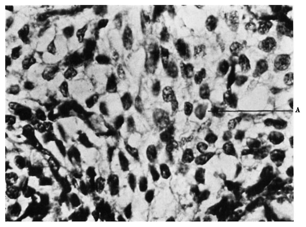 | 1,000 × 740 (118 KB) | Z8600021 (talk | contribs) | ==Plate XXVI== Fig. 4. Single erythrocyte (A) surrounded by mesodermic cells. 60 mm embryo. X 700. Fig. 5. On right is a “primaries ” of the first generation, with cartilage. On the left are “ primaries ” of the second generation. 68 mm embry... |
You cannot overwrite this file.
File usage
The following page uses this file: