File:Conklin 1905 plate11.jpg
Original file (1,521 × 2,000 pixels, file size: 710 KB, MIME type: image/jpeg)
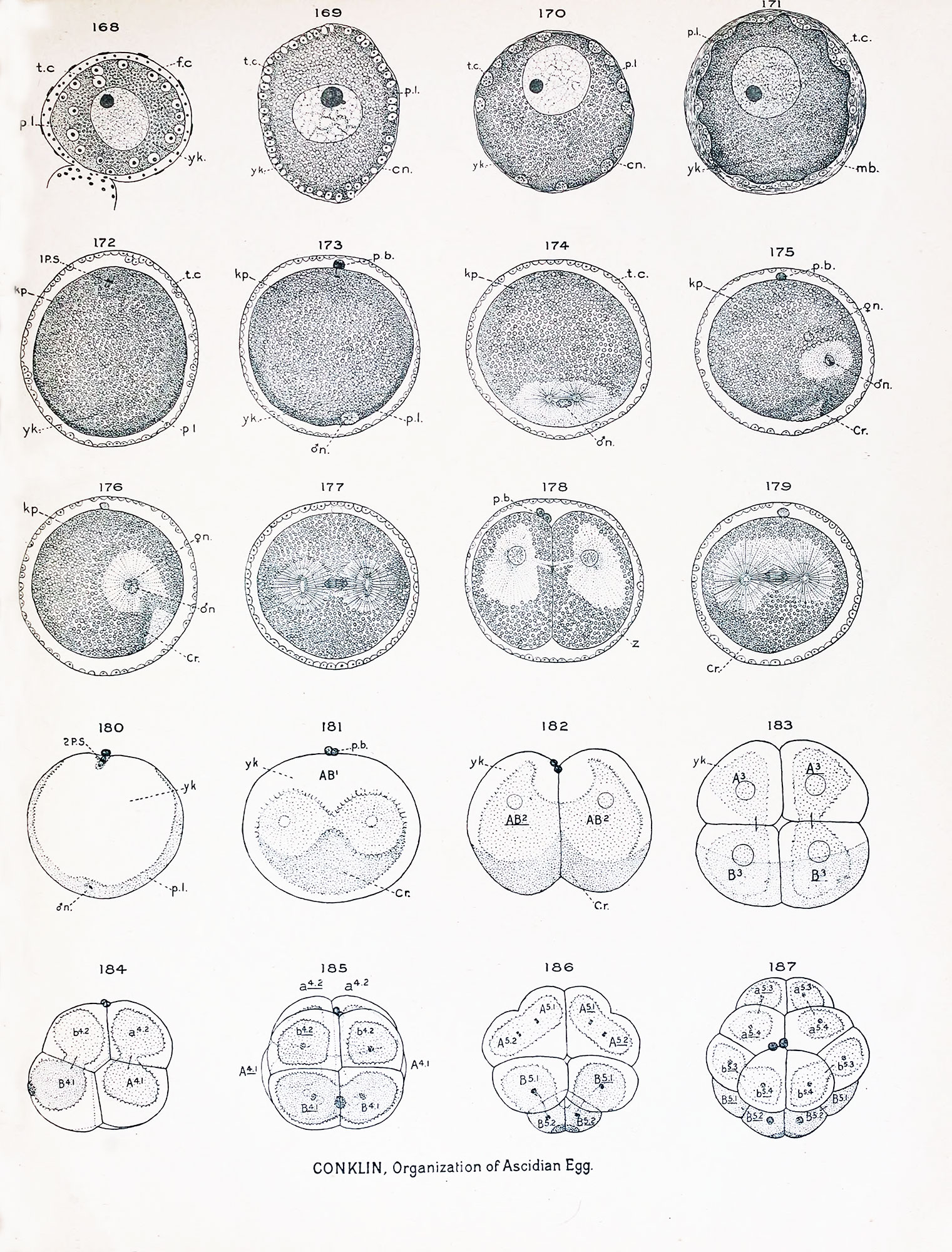
Plate XI Eggs of Ciona intestinalis - Ovocyte to Fourth Cleavage
Figs. 168-179 actual section*; figs. 180-187 whole eggs. Crescent substance and clear protoplasm stippled. Polar bodies are actually present where drawn.
Fig. 168. Half grown ovarian egg, showing test cells within the egg cytoplasm, which is composed of a layer of yolk surrounding the nucleus and a peripheral layer of clear protoplasm ; around the whole egg is a layer of small follicle cells which later develop into the very large conical cells which surround the chorion.
Fig. 169. Older ovarian egg, showing the area of the yolk increased and the test cells limited to the peripheral layer of protoplasm, which is sharply differentiated from the yolk.
Fig. 170. Still older ovarian egg, showing the test cells arranged in groups or "nests" at the periphery of the egg.
Fig. 171. Ripe ovarian egg ready to escape from the ovary, showing the extrusion of the test cells and the formation of a thick homogeneous membrane around the egg, which ultimately becomes the chorion.
Fig. 172. Free hut unfertilized egg which lias shrunken away from the chorion, showing the first maturation spindle, the peripheral layer of protoplasm collected over the lower pole of the egg and the achromatic substance of the germinal vesicle (kp) spread in a broad layer over the upper pole and around the central yolk.
Fig. 173. Extrusion of first polar body and entrance of spermatozoon ; the sperm nucleus lies in the peripheral layer surrounded by clear protoplasm in which astral rays are developing.
Fig. 174. Nearly equatorial section of the egg showing sperm nucleus and amphiaster at the posterior side of the egg; at the surface is the granular protoplasm of the peripheral layer.
Fig. 175. Section of egg in plane of first cleavage (median plane), showing the approach of the germ nuclei and the movement of the peripheral layer of protoplasm from the lower pole to the posterior side to form the crescent ; in the darkly staining substance of the crescent is a clear triangular area which corresponds to the clear area surrounding the sperm nucleus in fig. 173.
Fig. 176. Section of slightly older stage in plane of first cleavage, showing the union of the germ nuclei ; polar body out of the plane of section.
Fig. 177. Anaphase of first cleavage, showing the complete separateness of the nuclear and astral portions of the mitotic figure; the crescent substance shows at the ends of the spindle.
Fig, 178. Telophase of first cleavage, showing " zwischenkorper " (z), also the bending of spindle axis and shifting of cytoplasm and nuclei toward the animal pole.
Fig. 179. Anaphase of second cleavage, showing separateness of nuclear and astral portions of mitotic figure, also position of crescent on postero- dorsal side ; polar body out of the plane of section.
Fig. 180. Entire egg of Ciona, showing formation of second polar spindle, peripheral layer of protoplasm, sperm nucleus and aster.
Fig. 181. Anaphase of first cleavage viewed from posterior pole.
Fig. 182. Two-cell stage seen from posterior pole, cytoplasm and nuclei lie near animal pole, crescent near vegetal, fig. 183. Four-cell -stage seen from vegetal pole ; the crescent covers about one half the surface of the two posterior blastomeres.
Fig. 1S4. Eight cell-stage, left side, showing cap of deeply staining protoplasm at posterior pole of cells (B 4-1 ), which later goes into the posterior mesenchyme cells (B 7 ' J , fig. 200.)
Fig. 185. Eight cells, posterior view; spindles of the fourth cleavage present.
Fig. 186. Fourth cleavage, vegetal pole.
Fig. 187. Fourth cleavage, animal pole; 16 cell.-.
| Historic Disclaimer - information about historic embryology pages |
|---|
 |
- Conklin Figures: Fig 1-2 | Fig 3-6 | Fig 7-8 | Fig 9-12 | Fig 13-16 | Fig 17-20 | Fig 21-24 | Fig 25-26 | Fig 27-33 | Fig 34-35 | Plate I | Plate II | Plate III | Plate IV | Plate V | Plate VI | Plate VII | Plate VIII | Plate IX | Plate X | Plate XI | Plate XII
Reference
Conklin EG. The Organization and Cell-Lineage of the Ascidian Egg (1905) J. Acad., Nat. Sci. Phila. 13, 1.
Conklin 1905 TOC: I. The Ovarian Egg | II. Maturation and Fertilization | III. Orientation of Egg and Embryo | IV. Cell-Lineage | V. Later Development | VI. Comparisons with A.mphioxus and Amphibia | VII. The Organization of the Egg | Summary | Literature Cited | Explanation of Figures
Cite this page: Hill, M.A. (2024, April 27) Embryology Conklin 1905 plate11.jpg. Retrieved from https://embryology.med.unsw.edu.au/embryology/index.php/File:Conklin_1905_plate11.jpg
- © Dr Mark Hill 2024, UNSW Embryology ISBN: 978 0 7334 2609 4 - UNSW CRICOS Provider Code No. 00098G
File history
Click on a date/time to view the file as it appeared at that time.
| Date/Time | Thumbnail | Dimensions | User | Comment | |
|---|---|---|---|---|---|
| current | 16:40, 19 October 2016 | | 1,521 × 2,000 (710 KB) | Z8600021 (talk | contribs) |
You cannot overwrite this file.
File usage
The following 2 pages use this file: