File:Conklin 1905 plate08.jpg

Original file (1,521 × 2,000 pixels, file size: 572 KB, MIME type: image/jpeg)
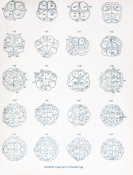
Plate VIII
Surface Views of Entire Eggs of Cynthia partita; Eight to Forty-four Cells.
Fig. 108. Eight-cell stage ; left side of egg ; showing spindles of third cleavage.
Fig. 109. Anterior view of 8-cell stage, showing cytoplasm most abundant in the animal pole cells,
and the yolk largely collected in the anterior cell of the vegetal hemisphere. Figs. 110, 111,112. Stages in the fourth cleavage; figs. 110 and 112 viewed from the animal pole, fig.
Ill from the. vegetal pole. Fig. 113. Telophase of fourth cleavage, vegetal poje view ; caps of deeply staining protoplasm lie at
the hinder borders of the small posterior cells (B 5,2 ). Figs. 114 and 115. Anterior and posterior views of the 16-cell -stage; fig. 115 showing caps of
deeply staining protoplasm at the posterior pole, which later go into the posterior mesenchyme cells (B' 6 , figs. 130, 131). Figs, lit! and 117. Ventral and dorsal views of a 20-cell stage, showing the cells at the vegetal pole
dividing before those at the animal pole. Fig. 118. Slightly older stage with some of the animal pole cells dividing. Figs. 119-123. Five views of one and the same egg; fig. 119, ventral; 120, dorsal; 121, anterior;
122, posterior; 123, right side; the latter shows in dotted outlines the great elongation
of the cells at the animal pole and the flattened shape of the cells at the vegetal pole ;
all the designations of cells in tig. 123 should be underscored ; 44 cells, 16 ectoderm, 10
endoderm, 10 mesoderm, 4 chorda and 4 neural plate cells. Figs. 124-129. Six different views of one and the same egg in the 44-cell stage showing the divisions
of the ectodermal cells and the second cells of the crescent (B 6 ' 4 ) ; when these divisions
are completed there will be 62 cells. Fig. 124, ventral; 125, dorsal; 126, anterior; 127, postero-dorsal.
File history
Click on a date/time to view the file as it appeared at that time.
| Date/Time | Thumbnail | Dimensions | User | Comment | |
|---|---|---|---|---|---|
| current | 16:19, 19 October 2016 |  | 1,521 × 2,000 (572 KB) | Z8600021 (talk | contribs) | ==Plate VIII== Surface Views of Entire Eggs of Cynthia partita; Eight to Forty-four Cells. Fig. 108. Eight-cell stage ; left side of egg ; showing spindles of third cleavage. Fig. 109. Anterior view of 8-cell stage, showing cytoplasm most abundant... |
You cannot overwrite this file.
File usage
The following 2 pages use this file: