File:Conklin 1905 plate02.jpg

Original file (1,521 × 2,000 pixels, file size: 282 KB, MIME type: image/jpeg)
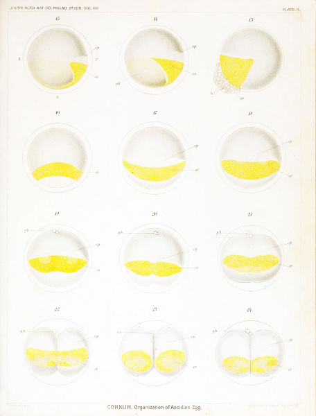
Plate II Living Eggs of Cynthia partita - First Cleavage
Figs. 13 and 14. Side views of egg, showing the formation of the crescent (cr.) from the yellow hemisphere ; in all the figures the animal pole is above, the vegetal pole below. Above the yellow crescent is an area of clear protoplasm (c. p.).
Fig. 15. Similar stage of another egg showing the aggregation of test cells over the crescent and the protrusion of the chorion at this place, an unusual phenomenon.
Fig. 16. Egg showing crescent, viewed nearly from the vegetal pule
Fig. 17. Another egg showing crescent and clear protoplasm, viewed from posterior pole; the animal pole is above, the vegetal below.
Figs. 18, 19, 20. Successive stages of the same egg drawn at intervals of about two minutes, showing the approaching division of the egg.
Figs. 21, 22, 23, 24. Succesive stages in the first cleavage of an egg, drawn at intervals of about two or three minutes. The egg is viewed from the posterior pole and shows the complicated forms taken by the yellow crescent during the division, also the enlargement of the area of clear protoplasm and its extension toward the animal pole.
| Historic Disclaimer - information about historic embryology pages |
|---|
 |
- Conklin Figures: Fig 1-2 | Fig 3-6 | Fig 7-8 | Fig 9-12 | Fig 13-16 | Fig 17-20 | Fig 21-24 | Fig 25-26 | Fig 27-33 | Fig 34-35 | Plate I | Plate II | Plate III | Plate IV | Plate V | Plate VI | Plate VII | Plate VIII | Plate IX | Plate X | Plate XI | Plate XII
Reference
Conklin EG. The Organization and Cell-Lineage of the Ascidian Egg (1905) J. Acad., Nat. Sci. Phila. 13, 1.
Conklin 1905 TOC: I. The Ovarian Egg | II. Maturation and Fertilization | III. Orientation of Egg and Embryo | IV. Cell-Lineage | V. Later Development | VI. Comparisons with A.mphioxus and Amphibia | VII. The Organization of the Egg | Summary | Literature Cited | Explanation of Figures
Cite this page: Hill, M.A. (2024, April 27) Embryology Conklin 1905 plate02.jpg. Retrieved from https://embryology.med.unsw.edu.au/embryology/index.php/File:Conklin_1905_plate02.jpg
- © Dr Mark Hill 2024, UNSW Embryology ISBN: 978 0 7334 2609 4 - UNSW CRICOS Provider Code No. 00098G
File history
Click on a date/time to view the file as it appeared at that time.
| Date/Time | Thumbnail | Dimensions | User | Comment | |
|---|---|---|---|---|---|
| current | 15:24, 19 October 2016 | | 1,521 × 2,000 (282 KB) | Z8600021 (talk | contribs) |
You cannot overwrite this file.
File usage
The following 2 pages use this file: