File:Congenital lobar emphysema.jpg

Original file (1,204 × 1,130 pixels, file size: 71 KB, MIME type: image/jpeg)
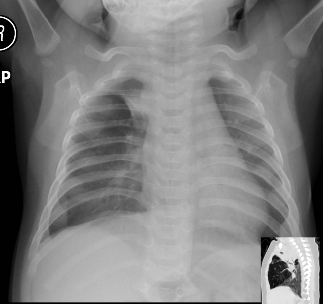
Congenital Lobar Emphysema (CLE)
Text below from Radiology Picture of Day
"The right middle lobe is hyperlucent, hyperexpanded with attenuated vascularity. It compresses adjacent parenchyma with RUL collapse.
CLE is a disorder affecting neonates and young infants and is usually associated with acute or subacute respiratory distress. Various bronchial and alveolar abnormalities can cause this disorder and in some cases the cause is unknown. The most common detected abnormality is absence or hypoplasia of cartilage rings of major and branch bronchi with resultant bronchial collapse during exhalation. This results in inhalational air entry but collapse of the narrow bronchial lumen during exhalation. The bronchial obstruction leads to progressive hyperinflation and air trapping, usually involving only one pulmonary lobe."
Image Source: http://www.radpod.org/2008/06/16/congenital-lobar-emphysema-2/
Credit: Dr Ahmed Haroun
This work is under a Creative Commons License
File history
Click on a date/time to view the file as it appeared at that time.
| Date/Time | Thumbnail | Dimensions | User | Comment | |
|---|---|---|---|---|---|
| current | 13:41, 24 August 2010 |  | 1,204 × 1,130 (71 KB) | S8600021 (talk | contribs) | ==Congenital Lobar Emphysema (CLE) == Text below from http://www.radpod.org/ Radiology Picture of Day "The right middle lobe is hyperlucent, hyperexpanded with attenuated vascularity. It compresses adjacent parenchyma with RUL collapse. CLE is a di |
You cannot overwrite this file.
File usage
The following page uses this file: