File:Colon histology 006.jpg: Difference between revisions
From Embryology
(Ano-Rectal Junction Crypt of Lieberkühn (transverse section) Intestinal gland Stain: van Gieson, H&E, trichrome Original File Name: ARecVG02.jpg Image Source: UWA Blue Histology Copyright Lutz Slomianka 1998-2009. The literary and artistic works () |
No edit summary |
||
| Line 1: | Line 1: | ||
Ano-Rectal Junction | ==Human Ano-Rectal Junction Histology== | ||
* alimentary canal, GIT, large intestine, rectum, mucosa, crypt of Lieberkühn, transverse section | |||
* Stain: van Gieson, H&E, trichrome | |||
{{Colon Histology Links}} | |||
Original File Name: ARecVG02.jpg | Original File Name: ARecVG02.jpg | ||
{{Blue Histology}} | |||
[[Category:Histology]] [[Category:Gastrointestinal Tract]] | [[Category:Histology]] [[Category:Gastrointestinal Tract]] | ||
Revision as of 07:06, 30 August 2011
Human Ano-Rectal Junction Histology
- alimentary canal, GIT, large intestine, rectum, mucosa, crypt of Lieberkühn, transverse section
- Stain: van Gieson, H&E, trichrome
- Colon Histology Links: Ano-Rectal Junction Overview Labeled | Colon Wall Labeled | Colon Mucosa Labeled | Colon Overview | Ano-Rectal Junction Overview | Intestinal Gland - longitudinal van Gieson | Intestinal Gland - transverse van Gieson | Intestinal Gland - longitudinal H&E | Intestinal Gland - transverse H&E | GIT Histology | Gastrointestinal Tract Development
Original File Name: ARecVG02.jpg
Links: Histology | Histology Stains | Blue Histology images copyright Lutz Slomianka 1998-2009. The literary and artistic works on the original Blue Histology website may be reproduced, adapted, published and distributed for non-commercial purposes. See also the page Histology Stains.
Cite this page: Hill, M.A. (2024, April 26) Embryology Colon histology 006.jpg. Retrieved from https://embryology.med.unsw.edu.au/embryology/index.php/File:Colon_histology_006.jpg
- © Dr Mark Hill 2024, UNSW Embryology ISBN: 978 0 7334 2609 4 - UNSW CRICOS Provider Code No. 00098G
File history
Click on a date/time to view the file as it appeared at that time.
| Date/Time | Thumbnail | Dimensions | User | Comment | |
|---|---|---|---|---|---|
| current | 09:37, 31 October 2011 |  | 400 × 533 (70 KB) | S8600021 (talk | contribs) | |
| 11:08, 2 November 2009 | 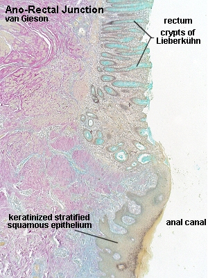 | 300 × 400 (113 KB) | S8600021 (talk | contribs) | Ano-Rectal Junction Crypt of Lieberkühn (transverse section) Intestinal gland Stain: van Gieson, H&E, trichrome Original File Name: ARecVG02.jpg Image Source: UWA Blue Histology Copyright Lutz Slomianka 1998-2009. The literary and artistic works ( |
You cannot overwrite this file.
File usage
The following 5 pages use this file: