File:Cochlea stria vascularis cartoon 01.jpg

Original file (874 × 1,897 pixels, file size: 338 KB, MIME type: image/jpeg)
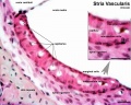
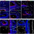
Human Cochlea Stria Vascularis
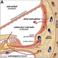
A cross-section through the adult cochlea. The cochlear duct (or scala media) is filled with endolymph containing a high [K+] that is maintained by the stria vascularis. Potassium recycling is postulated to either occur via the supporting cells of the organ of Corti and the epithelial lining of the outer sulcus (Claudius cells and root cells), or through the perilymph of the scala tympani.
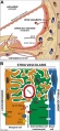
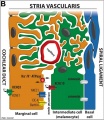
B anatomical (upper half) and compartmental (lower half) model of the adult stria vascularis showing the three cellular layers and depicting the location of potassium regulating channels. The stria vascularis is electrochemically isolated from neighboring structures by tight junctions (black bars).
- Cochlea Links: stria vascular histology | stria vascularis 1 | stria vascularis 2 | stria vascularis 3 | human vascularis development | Neural Crest Development | Inner Ear Development
stria vascular histology
stria vascular 1
stria vascular 2
stria vascular 3
vascularis development


Reference
Locher H, de Groot JC, van Iperen L, Huisman MA, Frijns JH & Chuva de Sousa Lopes SM. (2015). Development of the stria vascularis and potassium regulation in the human fetal cochlea: Insights into hereditary sensorineural hearing loss. Dev Neurobiol , 75, 1219-40. PMID: 25663387 DOI.
Copyright
© 2015 The Authors Developmental Neurobiology Published by Wiley Periodicals, Inc. This is an open access article under the terms of the Creative Commons Attribution-NonCommercial License, which permits use, distribution and reproduction in any medium, provided the original work is properly cited and is not used for commercial purposes.
Figure 1. relabelled with pubmed ID
Cite this page: Hill, M.A. (2024, April 27) Embryology Cochlea stria vascularis cartoon 01.jpg. Retrieved from https://embryology.med.unsw.edu.au/embryology/index.php/File:Cochlea_stria_vascularis_cartoon_01.jpg
- © Dr Mark Hill 2024, UNSW Embryology ISBN: 978 0 7334 2609 4 - UNSW CRICOS Provider Code No. 00098G
File history
Click on a date/time to view the file as it appeared at that time.
| Date/Time | Thumbnail | Dimensions | User | Comment | |
|---|---|---|---|---|---|
| current | 12:42, 11 April 2015 | | 874 × 1,897 (338 KB) | Z8600021 (talk | contribs) | ===Reference=== {pubmed>25663387</pubmed>| [http://onlinelibrary.wiley.com/doi/10.1002/dneu.22279/full Dev Neurobiol.] ====Copyright==== © 2015 The Authors Developmental Neurobiology Published by Wiley Periodicals, Inc. This is an open access articl... |
You cannot overwrite this file.
File usage
The following 7 pages use this file: