File:Chlamydia life cycle cartoon.jpg
Original file (910 × 800 pixels, file size: 112 KB, MIME type: image/jpeg)
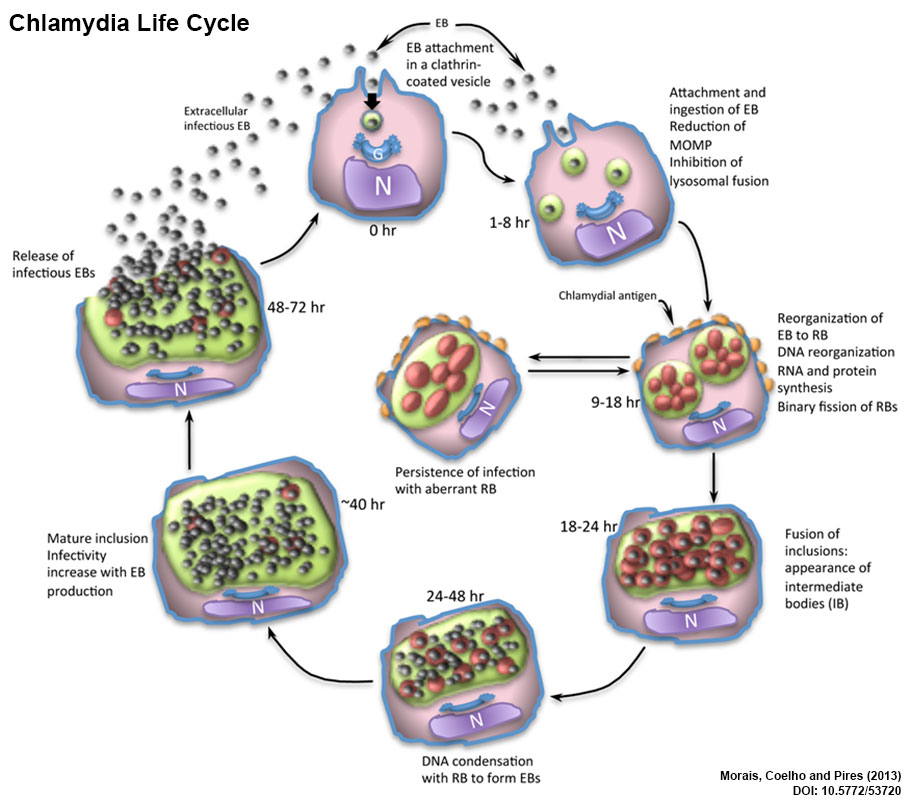
Chlamydia Life Cycle
Infection begins with the attachment of the elementary bodies (EB) to the surface of target epithelial cells. These cells promote a pseudopod formation to engulf the EB. Inside the cytoplasm this bacterium inhibits the fusion of the vesicle with the cell lysosomes. The nascent inclusion is accompanied by the transition from EBs to reticulate bodies (RB). Late in the cycle, RBs replicate by binary fission to generate both RBs and intermediate bodies (IB). At this stage, antigenic proteins are exposed into the cell surface. An elongated, aberrant RB could be formed at this time with an arrest on chlamydia cycle originating a persistent infection, or continuing the cycle. The various intracytoplasmic inclusions with bacterium inside, can also be fused in this phase, and the agent develop into intermediate bodies (IB), before DNA condensation and RB transformation into a newly EB. The mature inclusion increases in size with EB formation, until becoming infectious and released into the extracellular space to continue a new intracellular cycle.
N – nucleus; G – Golgi apparatus: EB – elementary bodies; RB – reticulate bodies; IB – intermediate bodies. (modified text from figure legend)
Reference
João Morais, Ana Cláudia Coelho and Maria dos Anjos Pires (2013). Psittacosis, Insights from Veterinary Medicine, Dr. Rita Payan Carreira (Ed.), InTech, DOI: 10.5772/53720. Available from: http://www.intechopen.com/books/insights-from-veterinary-medicine/psittacosis
Copyright
"Insights from Veterinary Medicine", book edited by Rita Payan-Carreira, ISBN 978-953-51-1005-7, Published: February 27, 2013 under CC BY 3.0 license. © The Author(s).
Cite this page: Hill, M.A. (2024, April 27) Embryology Chlamydia life cycle cartoon.jpg. Retrieved from https://embryology.med.unsw.edu.au/embryology/index.php/File:Chlamydia_life_cycle_cartoon.jpg
- © Dr Mark Hill 2024, UNSW Embryology ISBN: 978 0 7334 2609 4 - UNSW CRICOS Provider Code No. 00098G
File history
Click on a date/time to view the file as it appeared at that time.
| Date/Time | Thumbnail | Dimensions | User | Comment | |
|---|---|---|---|---|---|
| current | 13:59, 9 March 2017 | | 910 × 800 (112 KB) | Z8600021 (talk | contribs) |
You cannot overwrite this file.
File usage
The following page uses this file: