File:Cave1936 fig03.jpg
From Embryology

Size of this preview: 800 × 393 pixels. Other resolution: 1,000 × 491 pixels.
Original file (1,000 × 491 pixels, file size: 66 KB, MIME type: image/jpeg)
Fig. 3. The “transient appendix ”
(A) at the sixth week, (B) at the seventh week (after Kelly and Hurdon), (C) at the 20-mm. stage (after Gladstone).
Modern Notes: appendix | gastrointestinal abnormalities
Reference
Cave AJ. Appendix vermiformis duplex. (1936) J Anat. 70: 283-292. PMID 171045891
Cite this page: Hill, M.A. (2024, April 26) Embryology Cave1936 fig03.jpg. Retrieved from https://embryology.med.unsw.edu.au/embryology/index.php/File:Cave1936_fig03.jpg
- © Dr Mark Hill 2024, UNSW Embryology ISBN: 978 0 7334 2609 4 - UNSW CRICOS Provider Code No. 00098G
File history
Click on a date/time to view the file as it appeared at that time.
| Date/Time | Thumbnail | Dimensions | User | Comment | |
|---|---|---|---|---|---|
| current | 11:13, 26 May 2020 | | 1,000 × 491 (66 KB) | Z8600021 (talk | contribs) | |
| 11:10, 26 May 2020 | 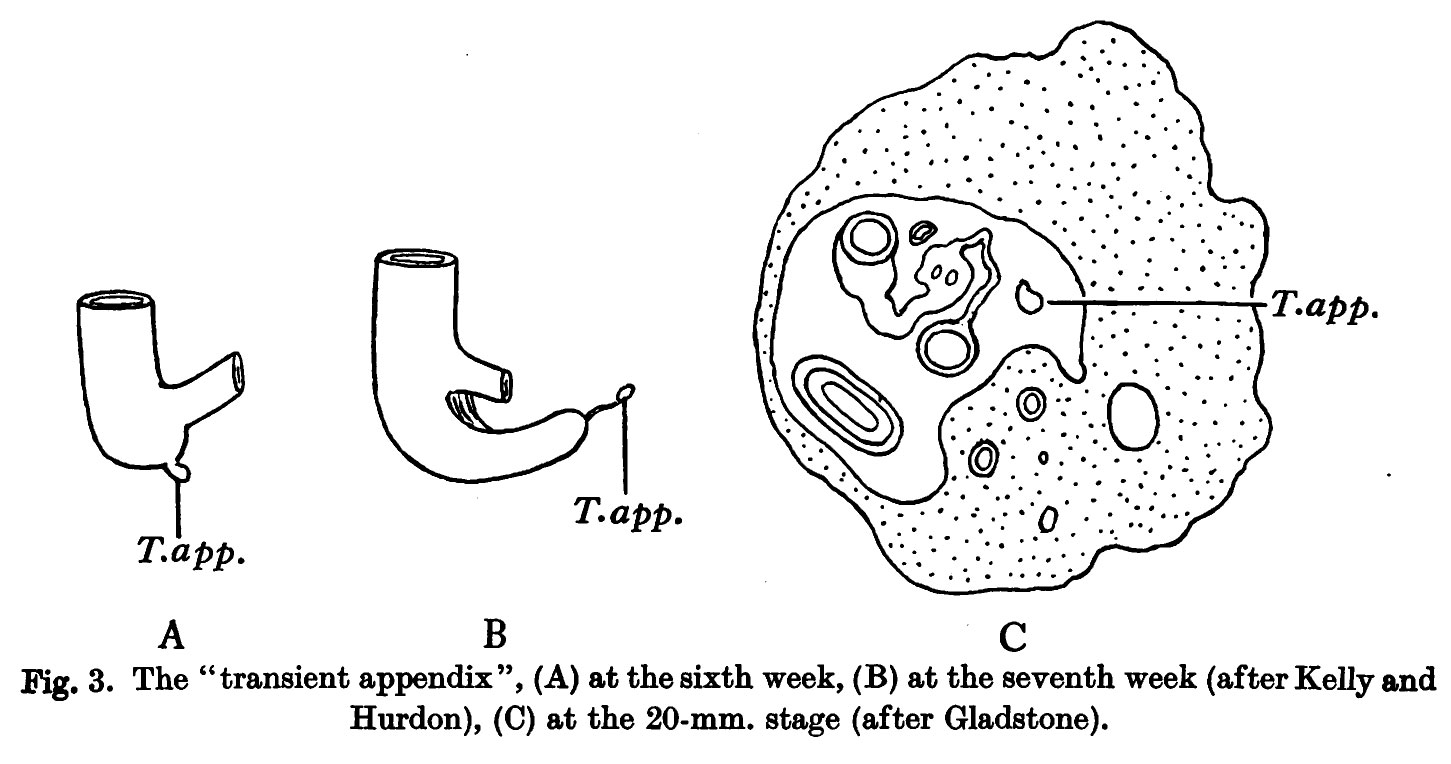 | 1,451 × 764 (134 KB) | Z8600021 (talk | contribs) | Fig. 3. The “transient appendix ”, (A) at the sixth week, (B) at the seventh week (after Kelly and Hurdon), (C) at the 20-mm. stage (after Gladstone). |
You cannot overwrite this file.
File usage
The following 2 pages use this file: