File:Catshark egg case stage 3.jpg
Catshark_egg_case_stage_3.jpg (710 × 390 pixels, file size: 76 KB, MIME type: image/jpeg)
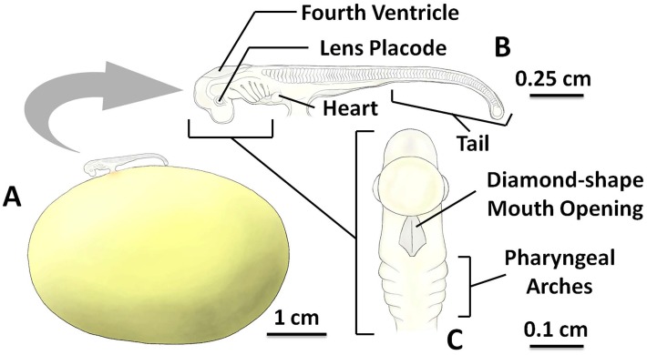
Catshark S. stellaris egg case at stage 3
A: The ellipsoid egg yolk mass and the associated embryo to scale.
B: The magnified embryo with indication of key morphological features (lateral view).
C: The diamond-shape mouth opening (ventral view). The key feature for stage 3 was the growth of the long tail.
See S3 File for original photographs of Fig 3 illustrations.
Reference
Musa SM, Czachur MV & Shiels HA. (2018). Oviparous elasmobranch development inside the egg case in 7 key stages. PLoS ONE , 13, e0206984. PMID: 30399186 DOI.
Copyright
© 2018 Musa et al. This is an open access article distributed under the terms of the Creative Commons Attribution License, which permits unrestricted use, distribution, and reproduction in any medium, provided the original author and source are credited.
Fig 3 Pone.0206984.g003.jpg
Cite this page: Hill, M.A. (2024, April 28) Embryology Catshark egg case stage 3.jpg. Retrieved from https://embryology.med.unsw.edu.au/embryology/index.php/File:Catshark_egg_case_stage_3.jpg
- © Dr Mark Hill 2024, UNSW Embryology ISBN: 978 0 7334 2609 4 - UNSW CRICOS Provider Code No. 00098G
File history
Click on a date/time to view the file as it appeared at that time.
| Date/Time | Thumbnail | Dimensions | User | Comment | |
|---|---|---|---|---|---|
| current | 10:13, 13 March 2019 |  | 710 × 390 (76 KB) | Z8600021 (talk | contribs) |
You cannot overwrite this file.
File usage
The following page uses this file:
