File:Cardiac muscle EM01.jpg
Original file (1,072 × 735 pixels, file size: 231 KB, MIME type: image/jpeg)
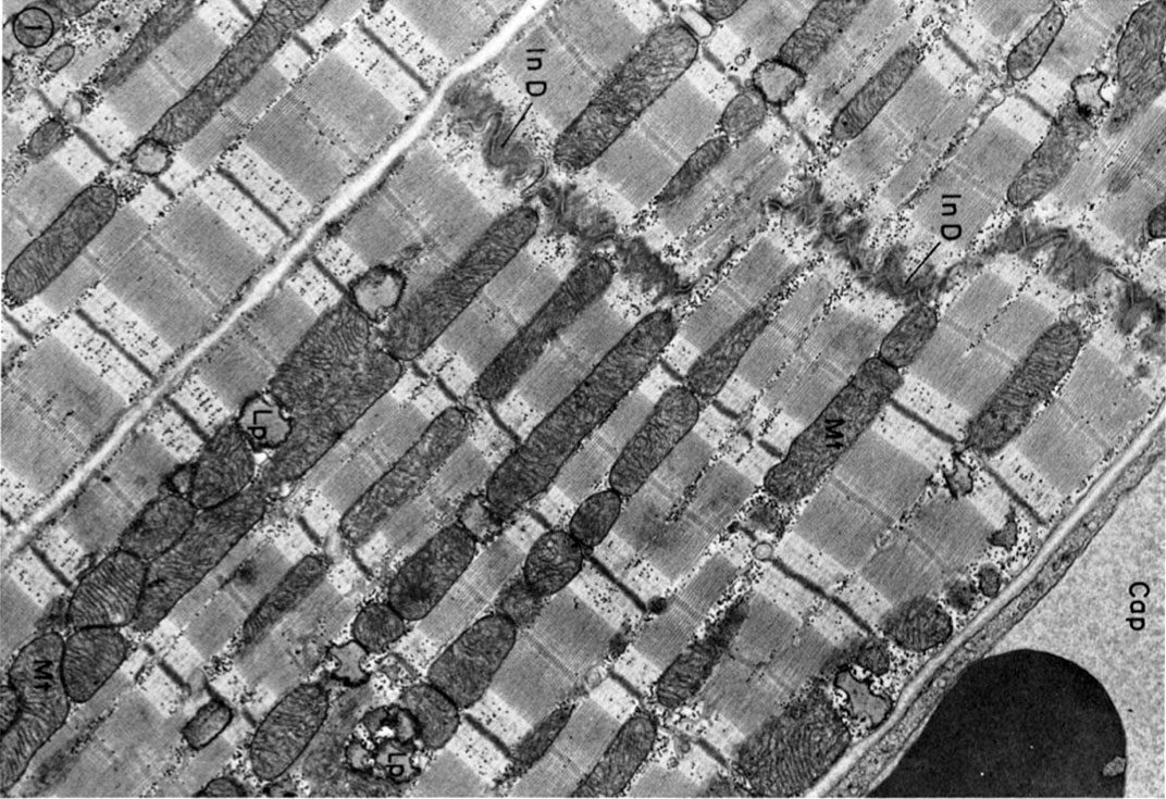
Cardiac Muscle EM
Electron micrograph of parts of three cat cardiac muscle fibers and an adjacent capillary (Cap) in longitudinal section. The two upper cells are joined end to end by a typical steplike intercalated disc (In D).Rows of mitochondria (Mt) appear to divide the contractile substance into myofibril-like units but, unlike the true myofibrils of skeletal muscle, these branch and rejoin and are quite variable in width. Lipid droplets (Lp) somewhat distorted in specimen preparation are found between the ends of the mitochondria. X 15,000.
Reference
<pubmed>4891913</pubmed>| PMC2107571
Copyright
Rockefeller University Press - Copyright Policy This article is distributed under the terms of an Attribution–Noncommercial–Share Alike–No Mirror Sites license for the first six months after the publication date (see http://www.jcb.org/misc/terms.shtml). After six months it is available under a Creative Commons License (Attribution–Noncommercial–Share Alike 4.0 Unported license, as described at https://creativecommons.org/licenses/by-nc-sa/4.0/ ). (More? Help:Copyright Tutorial)
File history
Click on a date/time to view the file as it appeared at that time.
| Date/Time | Thumbnail | Dimensions | User | Comment | |
|---|---|---|---|---|---|
| current | 11:52, 6 August 2012 | | 1,072 × 735 (231 KB) | Z8600021 (talk | contribs) | ==Cardiac Muscle EM== Electron micrograph of parts of three cat cardiac muscle fibers and an adjacent capillary (Cap) in longitudinal section. The two upper cells are joined end to end by a typical steplike intercalated disc (In D).Rows of mitochondria ( |
You cannot overwrite this file.
File usage
The following 5 pages use this file: