File:CameronMilligan1910 fig06-10.jpg

Original file (1,839 × 2,309 pixels, file size: 208 KB, MIME type: image/jpeg)
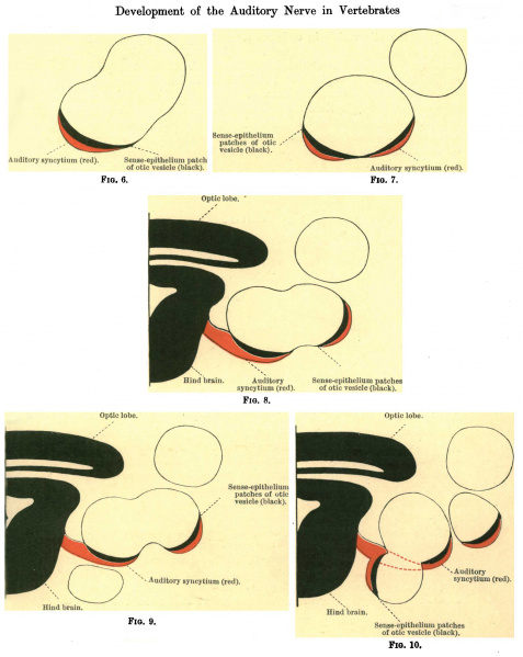
Figs. 6, 7, 8, 9 and 10 are camera lucida tracings of the otic vesicle and auditory syncytium of an embryo of Oyclopterus lumpus, a teleost
Oyclopterus lumpus - lumpsucker or lumpfish
They represent Nos. 96, 98, 100, 102, and 104 of a series of transverse sections, numbered from the cephalic end.
The syncytium, which is coloured red, will be observed in fig. 6 (section No. 96) to correspond in size with the thickened senseepithelium patch of the otic vesicle, with the cell-elements of which it is in intimate relationship.
In fig. 7 (section No. 98) the patch has divided into two areas, with a thinned portion of the vesicle wall between, and so likewise has the syncytium; the result being that the latter is still maintaining its close association with both.
In figs. 8 and 9 (sections Nos. 100 and 102) the two sense-epithelium patches are still seen, whilst the attachment of the syncytium to the wall of the hind brain is likewise clearly indicated.
In fig. 10 (section No. 104) the two patches have become completely separated, but the syncytium is preserving its intimate attachment to both. In the same figure a third patch has made its appearance on the semicircular canal in the lower part of the labyrinth, and it, in its turn, is in close association with an offshoot from the main syncytial mass.
From a further examination of embryos of Cyclopterus lumpus it was ascertained that these subsidiary sense-epithelium patches were all derivatives of the originally single area, with which the auditory syncytium was in continuity during the early developmental stages (fig. 11).
Reference
Cameron J. and Milligan W. The development of the auditory nerve in vertebrates. (1910) J Anat. Physiol. 44(2): 111-32. PubMed 17232833
Cite this page: Hill, M.A. (2024, April 26) Embryology CameronMilligan1910 fig06-10.jpg. Retrieved from https://embryology.med.unsw.edu.au/embryology/index.php/File:CameronMilligan1910_fig06-10.jpg
- © Dr Mark Hill 2024, UNSW Embryology ISBN: 978 0 7334 2609 4 - UNSW CRICOS Provider Code No. 00098G
File history
Click on a date/time to view the file as it appeared at that time.
| Date/Time | Thumbnail | Dimensions | User | Comment | |
|---|---|---|---|---|---|
| current | 17:02, 6 November 2017 | | 1,839 × 2,309 (208 KB) | Z8600021 (talk | contribs) |
You cannot overwrite this file.
File usage
The following page uses this file: