File:BurgosFawcett1955 fig13.jpg
From Embryology

Size of this preview: 427 × 599 pixels. Other resolution: 1,460 × 2,049 pixels.
Original file (1,460 × 2,049 pixels, file size: 501 KB, MIME type: image/jpeg)
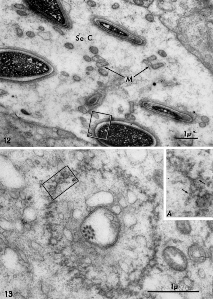
Fig. 13. A transverse section through the postnuclear region of an advanced spermatid showing in the center of the figure a cross—section of the tail flagellum surrounded by a pair of membranes. The larger ring in the spermatid cytoplasm is the so called caudal sheath or manchette. This is not a continuous membrane, as often described, but a cylindrical aggregation of delicate filaments. X 29,506. When examined at very high magnification these appear to be tubular. (See inset A.) X 65,000.
File history
Click on a date/time to view the file as it appeared at that time.
| Date/Time | Thumbnail | Dimensions | User | Comment | |
|---|---|---|---|---|---|
| current | 21:02, 30 May 2018 |  | 1,460 × 2,049 (501 KB) | Z8600021 (talk | contribs) | |
| 21:01, 30 May 2018 |  | 1,543 × 2,416 (523 KB) | Z8600021 (talk | contribs) | Fig. 13. A transverse section through the postnuclear region of an advanced spermatid showing in the center of the figure a cross—section of the tail flagellum surrounded by a pair of membranes. The larger ring in the spermatid cytoplasm is the so c... |
You cannot overwrite this file.
File usage
The following page uses this file: