File:Bulmer1957 plate01.jpg

Original file (1,280 × 1,808 pixels, file size: 353 KB, MIME type: image/jpeg)
Plate 1
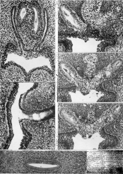
Fig. 1. 28 mm. embryo. Transverse section through the openings of the Wolffian ducts into the urogenital sinus. The sinus epithelium consists of basal deeply staining and superficial pale-staining cells, and the pale-staining cells extend for a short distance into the lower ends of the Wolffian ducts. The Miillerian ducts are in close relation with the Wolflian ducts, but a small mass of mesoderm separates them from the sinus. ( x 215.)
Fig. 2. 48 mm. foetus. Sagittal section through the opening of the right Wolfiian duct. The differentiation of the sinus epithelium in this region can be seen, and the extension of the pale-staining cells into the lower end of the Wolffian duct. ( x 215.)
Fig. 3. 65 mm. foetus. Transverse section through the genital cord. The Wolffian ducts are very small structures, on either side of the utero-vaginal canal. ( x 215.)
Fig. 4. 65 mm. foetus. Transverse section through the junction of Mullerian and sinus epithelia. A mass of darkly staining cells (d), arising from the dorsal wall of the sinus, separates the caudal end of the Mullerian epithelium (m) from the sinus lumen. On the right side the section passes below the bulk of the dorso-lateral projection, and the Wolffian duct is large. On the left the Wolffian duct is a small structure, applied to the dorsal aspect of the dorso-lateral projection (dp). (x 215.)
Fig. 5. 65 mm. foetus. Transverse section 16 p. caudal to Fig. 4. The Mullerian epithelium now forms a smaller mass dorsally, and a few of the darkly staining sinus cells separate it from the pale-staining sinus epithelium ventrally. ( x 215.)
Fig. 6. 65 mm. foetus. Transverse section 8 ,u caudal to Fig. 5. ( x 215.)
| Historic Disclaimer - information about historic embryology pages |
|---|
| Pages where the terms "Historic" (textbooks, papers, people, recommendations) appear on this site, and sections within pages where this disclaimer appears, indicate that the content and scientific understanding are specific to the time of publication. This means that while some scientific descriptions are still accurate, the terminology and interpretation of the developmental mechanisms reflect the understanding at the time of original publication and those of the preceding periods, these terms, interpretations and recommendations may not reflect our current scientific understanding. (More? Embryology History | Historic Embryology Papers) |
Reference
Bulmer D. The development of the human vagina. (1957) J. Anat. 91: 490-509.
Cite this page: Hill, M.A. (2024, April 28) Embryology Bulmer1957 plate01.jpg. Retrieved from https://embryology.med.unsw.edu.au/embryology/index.php/File:Bulmer1957_plate01.jpg
- © Dr Mark Hill 2024, UNSW Embryology ISBN: 978 0 7334 2609 4 - UNSW CRICOS Provider Code No. 00098G
File history
Click on a date/time to view the file as it appeared at that time.
| Date/Time | Thumbnail | Dimensions | User | Comment | |
|---|---|---|---|---|---|
| current | 18:43, 13 August 2018 |  | 1,280 × 1,808 (353 KB) | Z8600021 (talk | contribs) | |
| 18:42, 13 August 2018 |  | 1,751 × 2,580 (628 KB) | Z8600021 (talk | contribs) | ===Reference=== {{Ref-Bulmer1957}} |
You cannot overwrite this file.
File usage
The following page uses this file:
