File:Bryce1908 plate03.jpg
Original file (856 × 1,200 pixels, file size: 217 KB, MIME type: image/jpeg)
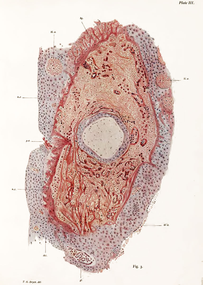
Plate 3.Section Through Blastocyst and Implantation Cavity
Fig. 3. Section Through Blastocyst and Implantation Cavity at the level of the point of entrance. x 100 1). dec.y decidua ; n,z., necrotic zone of decidua ; P.E., point of entrance ; gl., gland ; hl.v., maternal capillaries : hg., haemorrhage.
The cavity of the trophoblast is at this point tilled with niesoblast. The ct^totrophoUast appeal's as a blue-pink lamella ; the plasmodi -trophoblast as bands and threads of protoplasm stained of a dusky-red colour enclosing nuclei. The spaces in the plasniodium are occupied by maternal blood corpuscles. These come from maternal capillaries opened up Jis the implantation cavity enlarges. In the upper part (»f the section thei*e has been a considei-able haemorrhage into the decidua ; this has in part broken down the necrotic zone, and the mass of effused blocnl hsis imrtly torn up the plasmodial bands. The implantation cavity is lined by a necrotic zone of the decidua distinguished by its pink colour ; the unaltered parts of the decidua have a grey-blue tint. Within the necrotic zone are seen numbers of free cells. The glands of the decidua are dilated ; their epithelium is desquamating, and their lumen contains I'ed and white blood corpuscles. The blood-ve«sels are much dilated, more especially on the deep aspect of the decidua, where they form almost sinus-like spaces ; to the right and below, the endothelium of one wall of such a vessel is seen covering the decidua. the decidua is ci-owded with leucocytes. The point of entrance shows a depression filled with a mass of fibrin ; continuous with this and with the necrotic tissue, a fibrinous spur projects into the implantation cavity.
- Bryce 1908 Human Ovum: Plate 1 | Plate 2 | Plate 3 | Plate 4 | Plate 5 | Plate 6 | Plate 7 | Plate 8 | Plate 9 | Plate 10
| Historic Disclaimer - information about historic embryology pages |
|---|
 |
File history
Click on a date/time to view the file as it appeared at that time.
| Date/Time | Thumbnail | Dimensions | User | Comment | |
|---|---|---|---|---|---|
| current | 08:18, 3 November 2013 | | 856 × 1,200 (217 KB) | Z8600021 (talk | contribs) | ==Plate 3.== {{Historic Disclaimer}} |
You cannot overwrite this file.
File usage
The following page uses this file: