File:Bremer1914 plate05.jpg: Difference between revisions
No edit summary |
mNo edit summary |
||
| (2 intermediate revisions by the same user not shown) | |||
| Line 1: | Line 1: | ||
==Plate 5== | |||
'''6''' Grosser cmbryo, slide 3, row 5, sect. 3. From the edge of the body-stalk, to show surface mesothelium (Mes.) and mesothelial cord, leading from funnel (f.) to unlined space (a). Coe., coelom; h, smaller unlined space;c, endothelial cord. X circa 580. | |||
'''7''' Grosser embryo, slide 3, row 4, scct. 5. From the edge of the body-stalk, to show mesothelial cord leading to unlined space, containing corpuscles. c and d, mesothelial cords (see text); Coe., coelom. X circa 800. | |||
'''9''' Minot embryo, H.E.C. no. 825, sect. 25. Vesscl from the body-stalk, to show delamination of endothelium, and its extension as cords. Further descrip- tion in text. Coc., coelom; ~Z. U. , blood-vessel. X circa 800. | |||
'''10''' Minot embryo, H.E.C. no. 825, sect. 24. Angioblast net in the chorion, to show the irregular contour of the cord, suggesting amoeboid movements, and the extra-intimnl space even around the young branch. X circa 540. | |||
{{Bremer1914 figures}} | |||
Latest revision as of 21:50, 27 October 2015
Plate 5
6 Grosser cmbryo, slide 3, row 5, sect. 3. From the edge of the body-stalk, to show surface mesothelium (Mes.) and mesothelial cord, leading from funnel (f.) to unlined space (a). Coe., coelom; h, smaller unlined space;c, endothelial cord. X circa 580.
7 Grosser embryo, slide 3, row 4, scct. 5. From the edge of the body-stalk, to show mesothelial cord leading to unlined space, containing corpuscles. c and d, mesothelial cords (see text); Coe., coelom. X circa 800.
9 Minot embryo, H.E.C. no. 825, sect. 25. Vesscl from the body-stalk, to show delamination of endothelium, and its extension as cords. Further descrip- tion in text. Coc., coelom; ~Z. U. , blood-vessel. X circa 800.
10 Minot embryo, H.E.C. no. 825, sect. 24. Angioblast net in the chorion, to show the irregular contour of the cord, suggesting amoeboid movements, and the extra-intimnl space even around the young branch. X circa 540.
| Historic Disclaimer - information about historic embryology pages |
|---|
 |
- Links: Plate 1 | Plate 2 | Plate 3 | Plate 4 | Plate 5 | Bremer 1914 | Harvard Collection | Historic Embryology Papers
Reference
Bremer JL. The earliest blood-vessels in man. (1914) Amer. J Anat. 16(4): 447-475.
Cite this page: Hill, M.A. (2024, May 20) Embryology Bremer1914 plate05.jpg. Retrieved from https://embryology.med.unsw.edu.au/embryology/index.php/File:Bremer1914_plate05.jpg
- © Dr Mark Hill 2024, UNSW Embryology ISBN: 978 0 7334 2609 4 - UNSW CRICOS Provider Code No. 00098G
File history
Click on a date/time to view the file as it appeared at that time.
| Date/Time | Thumbnail | Dimensions | User | Comment | |
|---|---|---|---|---|---|
| current | 21:48, 27 October 2015 | 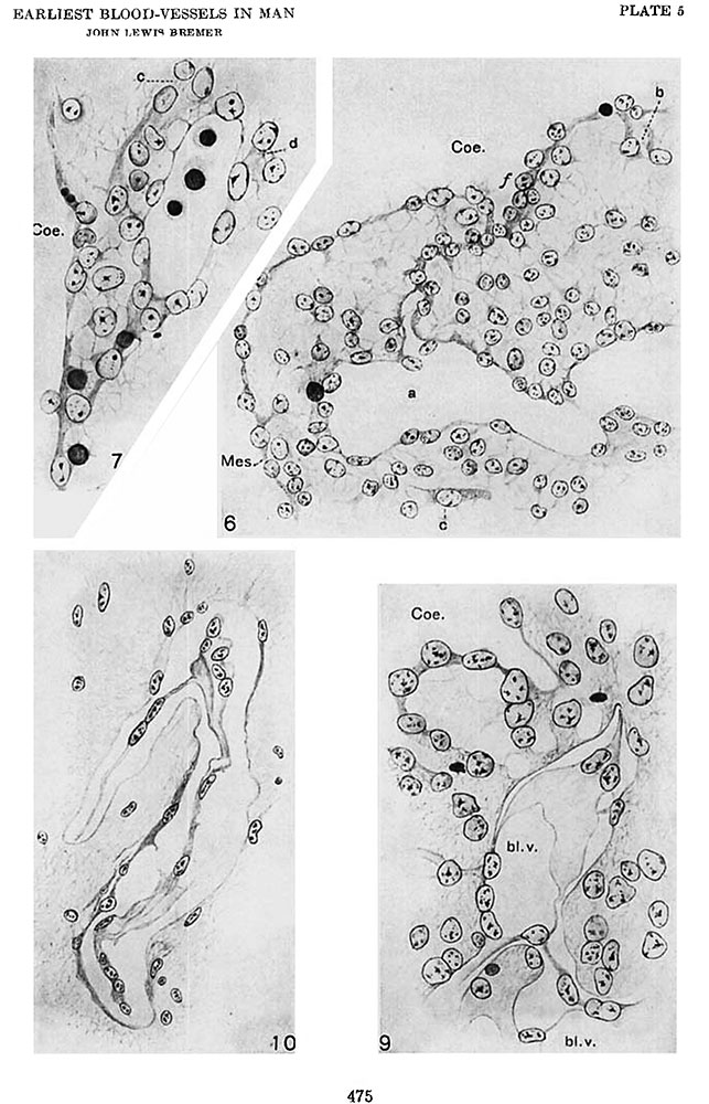 | 643 × 1,000 (144 KB) | Z8600021 (talk | contribs) |
You cannot overwrite this file.
File usage
The following 2 pages use this file: