File:Bremer1914 plate03.jpg: Difference between revisions
(==Plate 3== Grosser embryo, slide 3, row 3. Junction of body-stalk and yolk-sac, and connection of the two vascular nets, along the side oE the allantois, between the mesoderm and entoderm. All., cavity of allantois, looking into yolk-sac; Am., cavity...) |
m (→Plate 3) |
||
| Line 1: | Line 1: | ||
==Plate 3== | ==Plate 3== | ||
Grosser embryo, slide 3, row 3. Junction of body-stalk and yolk-sac, and connection of the two vascular nets, along the side | Grosser embryo, slide 3, row 3. Junction of body-stalk and yolk-sac, and connection of the two vascular nets, along the side of the allantois, between the mesoderm and entoderm. All., cavity of allantois, looking into yolk-sac; Am., cavity of amnion, which at one point is fused with the allantois;Ent., sheet of entoderm forming wall of yolk-sac and continued as allantois wall; Mes., sheet of mesothelium continued from yolk-sac to body-stalk; Ys., cavity of yolk-sac. X 400. | ||
Revision as of 21:39, 27 October 2015
Plate 3
Grosser embryo, slide 3, row 3. Junction of body-stalk and yolk-sac, and connection of the two vascular nets, along the side of the allantois, between the mesoderm and entoderm. All., cavity of allantois, looking into yolk-sac; Am., cavity of amnion, which at one point is fused with the allantois;Ent., sheet of entoderm forming wall of yolk-sac and continued as allantois wall; Mes., sheet of mesothelium continued from yolk-sac to body-stalk; Ys., cavity of yolk-sac. X 400.
| Historic Disclaimer - information about historic embryology pages |
|---|
 |
Reference
Bremer, JL. The Earliest Blood-Vessels in Man. (1914) Amer. J. Anat. Vol.16 No.4. 447-475.
Cite this page: Hill, M.A. (2024, May 8) Embryology Bremer1914 plate03.jpg. Retrieved from https://embryology.med.unsw.edu.au/embryology/index.php/File:Bremer1914_plate03.jpg
- © Dr Mark Hill 2024, UNSW Embryology ISBN: 978 0 7334 2609 4 - UNSW CRICOS Provider Code No. 00098G
File history
Click on a date/time to view the file as it appeared at that time.
| Date/Time | Thumbnail | Dimensions | User | Comment | |
|---|---|---|---|---|---|
| current | 21:38, 27 October 2015 | 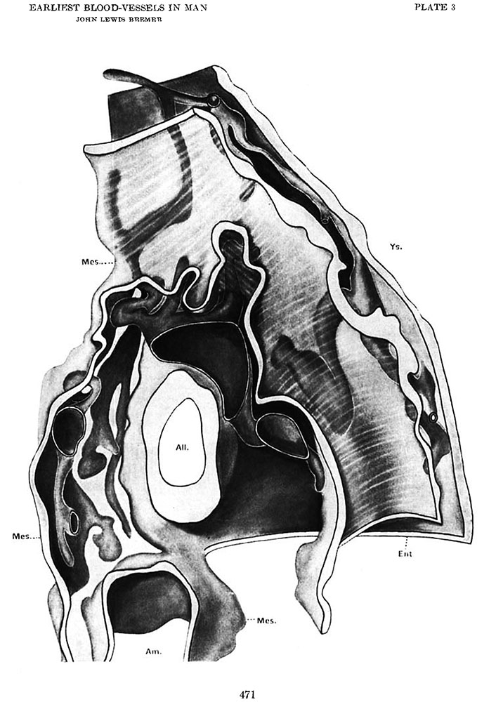 | 706 × 1,000 (127 KB) | Z8600021 (talk | contribs) | ==Plate 3== Grosser embryo, slide 3, row 3. Junction of body-stalk and yolk-sac, and connection of the two vascular nets, along the side oE the allantois, between the mesoderm and entoderm. All., cavity of allantois, looking into yolk-sac; Am., cavity... |
You cannot overwrite this file.
File usage
The following 2 pages use this file: