File:Braune 1877 plate 30 fig1.jpg
Original file (805 × 1,200 pixels, file size: 205 KB, MIME type: image/jpeg)
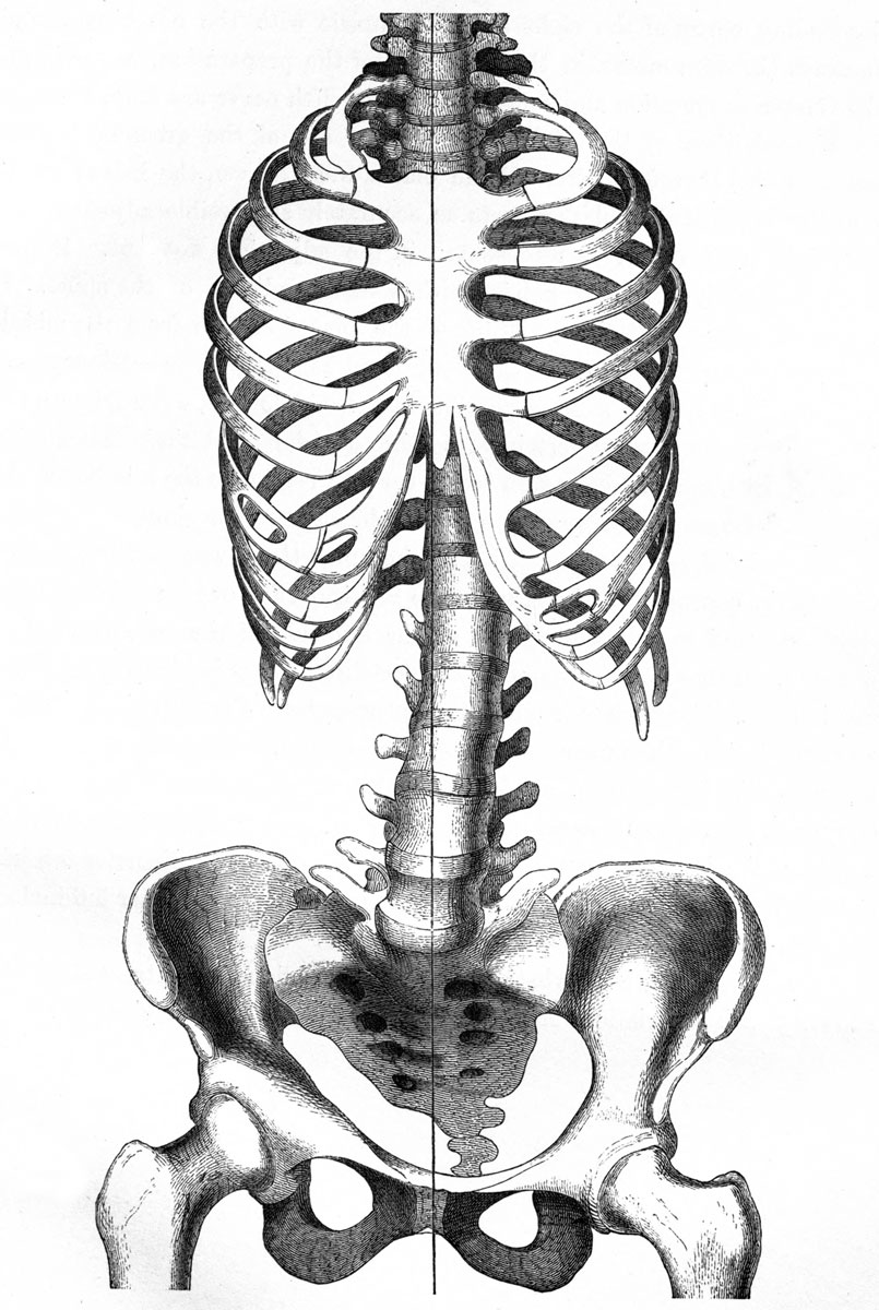
Plate 30 Fig. 1 Scoliosis
The relations of the skeleton, however, are of the greatest importance. I had therefore, after all the plates were drawn, the halves of the skeleton macerated, and the parts as accurately as possible adjusted with regard to each other, as represented in the adjoining woodcut. It presents a slightly scoliosed pelvis, with a like condition of the spine. It shows moreover that the deviation of the line of section from the middle line was not so considerable as the plate might suggest. The section passed through the pelvis, as near as possible in the middle line, externally and to the right of the lumbar vertebrge, meeting the dorsal at their articulation with the ribs, and passing again in the cervical region to the middle of the spinal column, and subsequently again to the right in the skull.
Beyond the scoliosed condition of the spine there was nothing worthy of remark, except that there were two cervical ribs, one complete on the right side, and a rudimentary one on the left side of the seventh cervical vertebra. There were seven cervical vertebrae, but only eleven dorsal and five lumbar. There was a rudimentary process from the fifth lumbar which was attached to the upper portion of the sacrum. The measurements of the pelvis in inches were as follows : The conjugata vera 3.8 in. (the conjugata at the narrowest points being 3.7) ; the right sacro-cotyloid 2.8 in. ; the left sacro-cotyloid 3.2 in. ; the transverse diameter 5.8 in. ; the left oblique diameter, 5.08 in., and the right oblique diameter 5.6 inches. The sacrum was 4.5 in. deep and 4.8 in. broad.
- Links: scoliosis | axial skeleton
| Embryology - 27 Apr 2024 |
|---|
| Google Translate - select your language from the list shown below (this will open a new external page) |
|
العربية | català | 中文 | 中國傳統的 | français | Deutsche | עִברִית | हिंदी | bahasa Indonesia | italiano | 日本語 | 한국어 | မြန်မာ | Pilipino | Polskie | português | ਪੰਜਾਬੀ ਦੇ | Română | русский | Español | Swahili | Svensk | ไทย | Türkçe | اردو | ייִדיש | Tiếng Việt These external translations are automated and may not be accurate. (More? About Translations) |
Braune W. An atlas of topographical anatomy after plane sections of frozen bodies. (1877) Trans. by Edward Bellamy. Philadelphia: Lindsay and Blakiston.
- Plates: 1. Male - Sagittal body | 2. Female - Sagittal body | 3. Obliquely transverse head | 4. Transverse internal ear | 5. Transverse head | 6. Transverse neck | 7. Transverse neck and shoulders | 8. Transverse level first dorsal vertebra | 9. Transverse thorax level of third dorsal vertebra | 10. Transverse level aortic arch and fourth dorsal vertebra | 11. Transverse level of the bulbus aortae and sixth dorsal vertebra | 12. Transverse level of mitral valve and eighth dorsal vertebra | 13. Transverse level of heart apex and ninth dorsal vertebra | 14. Transverse liver stomach spleen at level of eleventh dorsal vertebra | 15. Transverse pancreas and kidneys at level of L1 vertebra | 16. Transverse through transverse colon at level of intervertebral space between L3 L4 vertebra | 17. Transverse pelvis at level of head of thigh bone | 18. Transverse male pelvis | 19. knee and right foot | 20. Transverse thigh | 21. Transverse left thigh | 22. Transverse lower left thigh and knee | 23. Transverse upper and middle left leg | 24. Transverse lower left leg | 25. Male - Frontal thorax | 26. Elbow-joint hand and third finger | 27. Transverse left arm | 28. Transverse left fore-arm | 29. Sagittal female pregnancy | 30. Sagittal female pregnancy | 31. Sagittal female at term
| Historic Disclaimer - information about historic embryology pages |
|---|
 |
File history
Click on a date/time to view the file as it appeared at that time.
| Date/Time | Thumbnail | Dimensions | User | Comment | |
|---|---|---|---|---|---|
| current | 15:04, 31 October 2012 | | 805 × 1,200 (205 KB) | Z8600021 (talk | contribs) | {{Braune 1877 header}} |
You cannot overwrite this file.