File:Braune 1877 plate 3.jpg

Original file (1,012 × 1,200 pixels, file size: 242 KB, MIME type: image/jpeg)
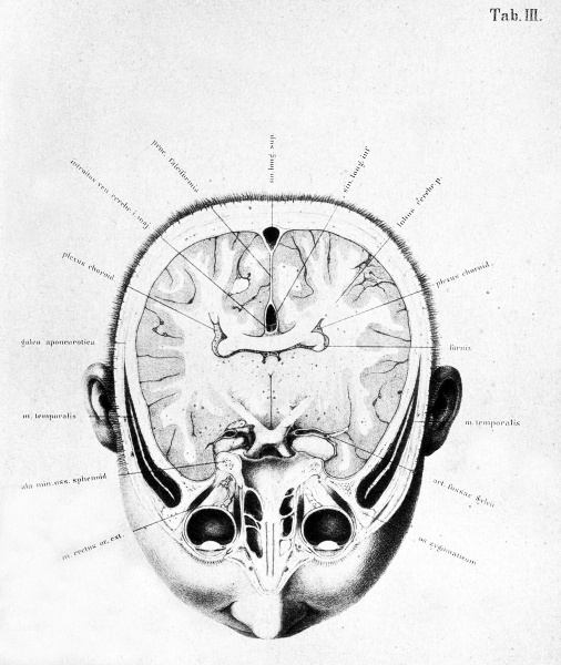
Plate 3 Obliquely Transverse Head
Only so much is certain, that the optic tract rises up from the chiasma to the corpora quadrigemina considerably more vertically than the optic nerve does to the optic foramen. I was therefore unable to expose the optic nerve thoroughly throughout its entire length, but was obliged to supplement the section by taking off a thin slice of the anterior lobe in order to completely expose the chiasma. Again, a thin layer of fat was removed from the orbit so as to show the entire breadth of the nerve, as the line of section had just missed its upper edge.
It must be further explained that, although the external form of the globe be established, the relations of the lens and iris must be rendered after further sections. The fine dust which even such a thin saw produces was very difficult to remove without causing a change in the relative position of the individual organs of the eye. I therefore froze fresh orbits, sawed through the bones, and then continued the section with a razor. In all cases the eye was thoroughly injected with Thiersch's carmine and glue preparation, in order to give the globe its original expansion. The injection was made from the ophthalmic artery, and in the entirely divided skull which forms PI. Ill, the carotid artery and jugular vein were completely injected with different colours.
It will be noticed from the relations of the brain that the plane of section is obliquely upwards and backwards. In front, owing to the removal of the thin lamina of the anterior lobe, a small portion of the floor of the skull in the region of the crista galli is seen. Behind it are the optic chiasma with a small piece of the optic tract cut obliquely, and further back is a section of the gyrus fornicatus, the superior processes of which lose themselves in the white substance of the cerebrum, and show the small bundles of fibres, the beak-shaped processes which belong to the fornix below.
Externally are the choroid plexuses of the descending cornua. Beneath the white substance of the corpus callosum is a fissure bounded laterally by the optic thalamus and filled up with vessels, and in the middle of it is the pineal body. In this space also is the pia mater passing beneath the corpus callosum to the central portion of the cerebrum. In the middle are the lumina of two large vessels belonging to the great internal veins of the brain, the vense magnse Galeni. These, when followed down with the sound under the splenium to the great veins behind the corpus callosum, and the commencement of the straight sinus, are found to debouch by the two veins here shown. The falx cerebri unites the inferior longitudinal sinus which opens into the straight sinus, with the superior longitudinal which lies further back.
| Embryology - 27 Apr 2024 |
|---|
| Google Translate - select your language from the list shown below (this will open a new external page) |
|
العربية | català | 中文 | 中國傳統的 | français | Deutsche | עִברִית | हिंदी | bahasa Indonesia | italiano | 日本語 | 한국어 | မြန်မာ | Pilipino | Polskie | português | ਪੰਜਾਬੀ ਦੇ | Română | русский | Español | Swahili | Svensk | ไทย | Türkçe | اردو | ייִדיש | Tiếng Việt These external translations are automated and may not be accurate. (More? About Translations) |
Braune W. An atlas of topographical anatomy after plane sections of frozen bodies. (1877) Trans. by Edward Bellamy. Philadelphia: Lindsay and Blakiston.
- Plates: 1. Male - Sagittal body | 2. Female - Sagittal body | 3. Obliquely transverse head | 4. Transverse internal ear | 5. Transverse head | 6. Transverse neck | 7. Transverse neck and shoulders | 8. Transverse level first dorsal vertebra | 9. Transverse thorax level of third dorsal vertebra | 10. Transverse level aortic arch and fourth dorsal vertebra | 11. Transverse level of the bulbus aortae and sixth dorsal vertebra | 12. Transverse level of mitral valve and eighth dorsal vertebra | 13. Transverse level of heart apex and ninth dorsal vertebra | 14. Transverse liver stomach spleen at level of eleventh dorsal vertebra | 15. Transverse pancreas and kidneys at level of L1 vertebra | 16. Transverse through transverse colon at level of intervertebral space between L3 L4 vertebra | 17. Transverse pelvis at level of head of thigh bone | 18. Transverse male pelvis | 19. knee and right foot | 20. Transverse thigh | 21. Transverse left thigh | 22. Transverse lower left thigh and knee | 23. Transverse upper and middle left leg | 24. Transverse lower left leg | 25. Male - Frontal thorax | 26. Elbow-joint hand and third finger | 27. Transverse left arm | 28. Transverse left fore-arm | 29. Sagittal female pregnancy | 30. Sagittal female pregnancy | 31. Sagittal female at term
| Historic Disclaimer - information about historic embryology pages |
|---|
 |
File history
Click on a date/time to view the file as it appeared at that time.
| Date/Time | Thumbnail | Dimensions | User | Comment | |
|---|---|---|---|---|---|
| current | 14:21, 31 October 2012 | | 1,012 × 1,200 (242 KB) | Z8600021 (talk | contribs) | {{Braune 1877 header}} |
You cannot overwrite this file.
File usage
The following 2 pages use this file: