File:Braune 1877 plate 29B.jpg

Original file (868 × 1,200 pixels, file size: 322 KB, MIME type: image/jpeg)
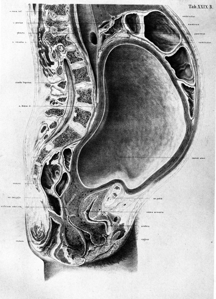
Plate 29. Sagittal Female Pregnancy (lower)
THE body from which this preparation was made was quite recent, twenty-five years of age, in the last month of pregnancy, and received in the condition of rigor mortis.
The foetus, which was divided in the section of the body, was subsequently restored to its original condition, so as to afford a representation of its former position in the uterus. I chiselled out the foetus and the liquor amnii from the left side of the body, and moistened the surface of the section of the uterus, and then froze it on the right side. The portion now lying in the right half of the uterus remained then for the purposes of representation as an untouched foetus. The left half of the uterus and its appendages, after the removal of the rest of the liquor amnii, was represented as empty. The foetus, which was in the second position of the head, was a well-formed female. The vulva were closed and the nails well developed. Its entire length was about twenty-three inches, its weight without the cord about six pounds. The cord was divided, and passed to the placenta between the head and right arm, the placenta being placed downwards and on the right side of the uterus. The child, as the plate shows, lay mostly in the right half of the uterus. In the section more than the right half of the head which was sawn obliquely, was removed. Moreover, the left arm and a portion of the right shoulder were divided longitudinally, and the forearm being placed at right angles with it, transversely, as well as a portion of the right leg, which extended towards the left side. The left knee was moreover grazed by the saw. The back and belly lay in the right half of the uterus, and the greater portion of the liquor amnii remained in the left. As the relations of this oblique section of the foetus offer points of no peculiar interest, I have refrained from reproducing the corresponding plate of the large atlas in this small edition.
The uterus is so folded over the symphysis that its anterior wall forms a kind of sac, indicating a condition of relaxation. The numerous large veins in its tissue are shown in the plate in the wall as simple strokes, their lumina becoming recognisable only when their walls were separated from each other; they appear patent, however, in the vaginal portion of the uterus and in the vagina itself. The vaginal portion of the uterus is proportionately deep, and for the most part lies in the left half of the body, the section having passed through its right half and opened merely the first portion of the cervix, as shown in Plate XXIX u. It was filled with viscid mucus and opened into the cavity of the uterus, about one fifth of an inch below the plane of section, so that its upper half could not be seen. The length of the vagina at this period of pregnancy makes it probable that the woman was not a primipara, notwithstanding that there were no cicatrices on the abdominal parietes, and the os internum was so narrow that only a very small sound could pass it. The number of veins met with in the right half of the vagina and their swollen condition is remarkable, and their lumina are peculiarly well seen in the left half of the preparation, Plate XXIX B. The falling in of the vaginal portion of the uterus is remarkable, considering the empty contracted condition of the bladder. The latter has slipped down bodily from the inner surface of the symphysis, and is so completely displaced that the course of the urethra has become bent at an angle. The external os lies in the hollow of the under border of the symphysis, although, according to Moreau, it corresponds at the end of pregnancy with the level of the upper border of the symphysis, and is still higher according to Schultze.
The level of the fundus corresponds nearly with the under border of the first lumbar vertebra; a more accurate definition cannot be given, as the highest point of the uterus was not included in the section, as it inclined more to the right side. This is almost the level given by Moreau, and according to the measurements of Schultze (' Wandtafeln,' taf. vi), it would appear to be the second lumbar vertebra. As the parts in the meanwhile began to thaw, a more accurate measurement in this particular could not be made.
| Embryology - 27 Apr 2024 |
|---|
| Google Translate - select your language from the list shown below (this will open a new external page) |
|
العربية | català | 中文 | 中國傳統的 | français | Deutsche | עִברִית | हिंदी | bahasa Indonesia | italiano | 日本語 | 한국어 | မြန်မာ | Pilipino | Polskie | português | ਪੰਜਾਬੀ ਦੇ | Română | русский | Español | Swahili | Svensk | ไทย | Türkçe | اردو | ייִדיש | Tiếng Việt These external translations are automated and may not be accurate. (More? About Translations) |
Braune W. An atlas of topographical anatomy after plane sections of frozen bodies. (1877) Trans. by Edward Bellamy. Philadelphia: Lindsay and Blakiston.
- Plates: 1. Male - Sagittal body | 2. Female - Sagittal body | 3. Obliquely transverse head | 4. Transverse internal ear | 5. Transverse head | 6. Transverse neck | 7. Transverse neck and shoulders | 8. Transverse level first dorsal vertebra | 9. Transverse thorax level of third dorsal vertebra | 10. Transverse level aortic arch and fourth dorsal vertebra | 11. Transverse level of the bulbus aortae and sixth dorsal vertebra | 12. Transverse level of mitral valve and eighth dorsal vertebra | 13. Transverse level of heart apex and ninth dorsal vertebra | 14. Transverse liver stomach spleen at level of eleventh dorsal vertebra | 15. Transverse pancreas and kidneys at level of L1 vertebra | 16. Transverse through transverse colon at level of intervertebral space between L3 L4 vertebra | 17. Transverse pelvis at level of head of thigh bone | 18. Transverse male pelvis | 19. knee and right foot | 20. Transverse thigh | 21. Transverse left thigh | 22. Transverse lower left thigh and knee | 23. Transverse upper and middle left leg | 24. Transverse lower left leg | 25. Male - Frontal thorax | 26. Elbow-joint hand and third finger | 27. Transverse left arm | 28. Transverse left fore-arm | 29. Sagittal female pregnancy | 30. Sagittal female pregnancy | 31. Sagittal female at term
| Historic Disclaimer - information about historic embryology pages |
|---|
| Pages where the terms "Historic" (textbooks, papers, people, recommendations) appear on this site, and sections within pages where this disclaimer appears, indicate that the content and scientific understanding are specific to the time of publication. This means that while some scientific descriptions are still accurate, the terminology and interpretation of the developmental mechanisms reflect the understanding at the time of original publication and those of the preceding periods, these terms, interpretations and recommendations may not reflect our current scientific understanding. (More? Embryology History | Historic Embryology Papers) |
File history
Click on a date/time to view the file as it appeared at that time.
| Date/Time | Thumbnail | Dimensions | User | Comment | |
|---|---|---|---|---|---|
| current | 14:45, 31 October 2012 |  | 868 × 1,200 (322 KB) | Z8600021 (talk | contribs) | {{Braune 1877 header}} |
You cannot overwrite this file.
File usage
The following 2 pages use this file:
