File:Brambell1927a plate29.jpg
Original file (1,753 × 2,464 pixels, file size: 290 KB, MIME type: image/jpeg)
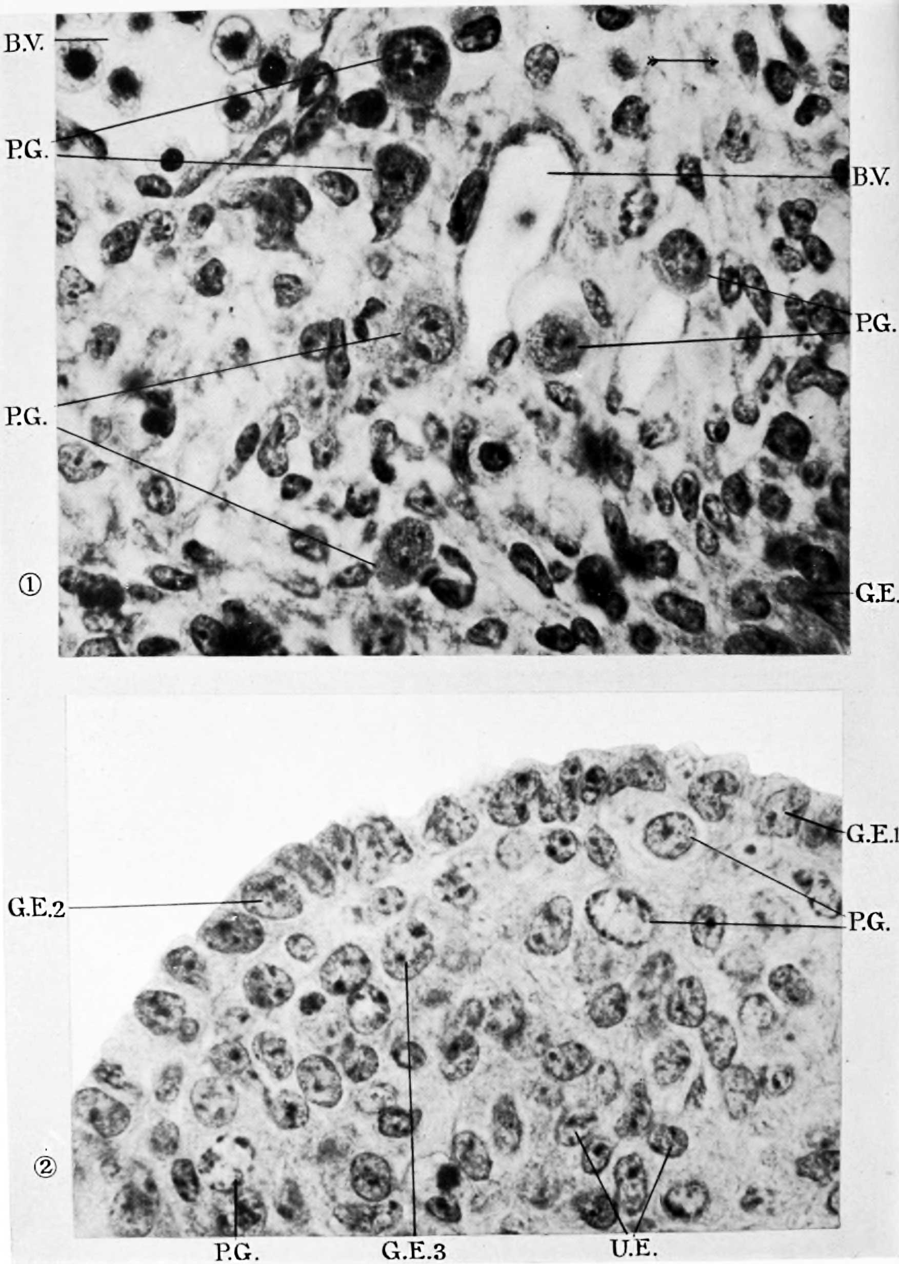
Plate 29
Fig. l. Six primordial germ-cells in the body-wall above the germinal ridge of a 10-day embryo. x 1000.
Fig. 2. Germinal ridge of a 10-day embryo showing cells proliferated from the epithelium in the undifierentiated condition and in various stages of differentiation into germ-cells. x 1000.
| Historic Disclaimer - information about historic embryology pages |
|---|
 |
Reference
Brambell FWR. The development and morphology of the gonads of the mouse. Part I. The morphogenesis of the indifferent gonad and of the ovary. (1927) 101: 391-407.
Cite this page: Hill, M.A. (2024, April 28) Embryology Brambell1927a plate29.jpg. Retrieved from https://embryology.med.unsw.edu.au/embryology/index.php/File:Brambell1927a_plate29.jpg
- © Dr Mark Hill 2024, UNSW Embryology ISBN: 978 0 7334 2609 4 - UNSW CRICOS Provider Code No. 00098G
File history
Click on a date/time to view the file as it appeared at that time.
| Date/Time | Thumbnail | Dimensions | User | Comment | |
|---|---|---|---|---|---|
| current | 21:50, 26 April 2018 | | 1,753 × 2,464 (290 KB) | Z8600021 (talk | contribs) |
You cannot overwrite this file.
File usage
The following page uses this file: