File:Bradley1908 fig10.jpg
From Embryology

Size of this preview: 800 × 581 pixels. Other resolution: 1,280 × 930 pixels.
Original file (1,280 × 930 pixels, file size: 244 KB, MIME type: image/jpeg)
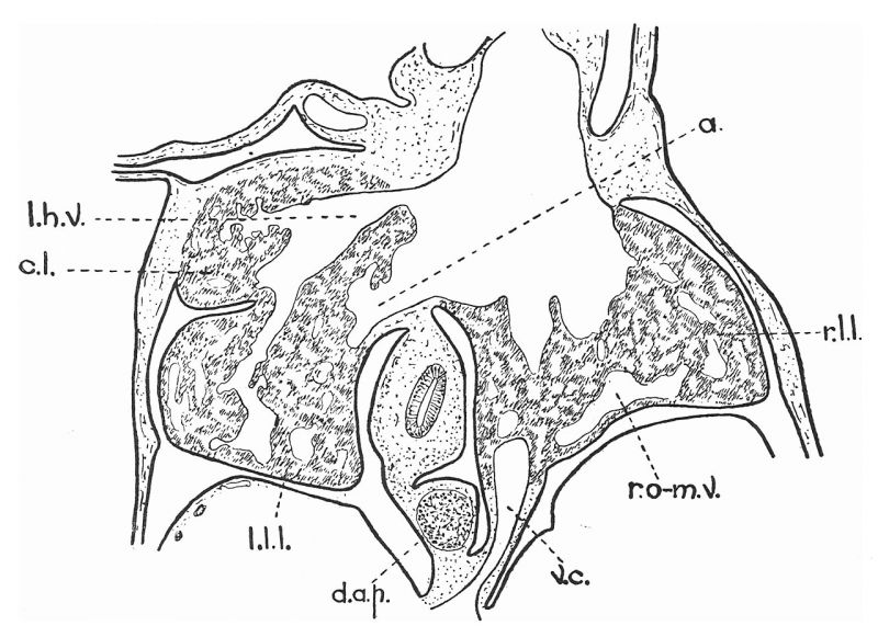
Fig. 10. Camera-lucida drawing of section of liver, etc., of 8-mm. pig embryo
V.c., Vena cava; a@, a small, short vein which ultimately becomes the ramus angularis. Figs. 10 to 14, inclusive, are of sections taken in order from the dorsal to the ventral part of the embryo.
Reference
Bradley OC. A contribution to the morphology and development of the mammalian liver. (1908) J Anat. 43: 1-42. PMID 17232788
Cite this page: Hill, M.A. (2024, April 27) Embryology Bradley1908 fig10.jpg. Retrieved from https://embryology.med.unsw.edu.au/embryology/index.php/File:Bradley1908_fig10.jpg
- © Dr Mark Hill 2024, UNSW Embryology ISBN: 978 0 7334 2609 4 - UNSW CRICOS Provider Code No. 00098G
File history
Click on a date/time to view the file as it appeared at that time.
| Date/Time | Thumbnail | Dimensions | User | Comment | |
|---|---|---|---|---|---|
| current | 21:45, 21 November 2019 | | 1,280 × 930 (244 KB) | Z8600021 (talk | contribs) | |
| 21:43, 21 November 2019 | 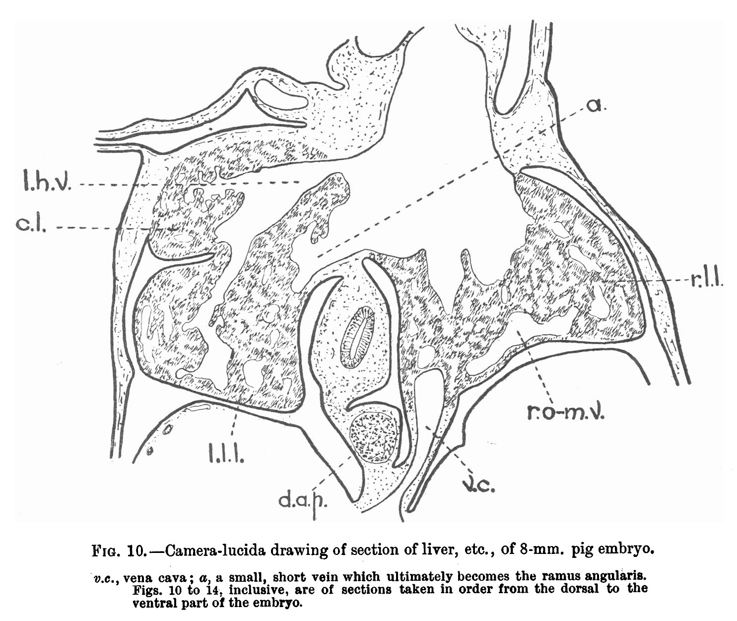 | 1,485 × 1,250 (305 KB) | Z8600021 (talk | contribs) |
You cannot overwrite this file.
File usage
The following 2 pages use this file: