File:Bradley1904a plate48.jpg
Original file (1,280 × 1,704 pixels, file size: 231 KB, MIME type: image/jpeg)
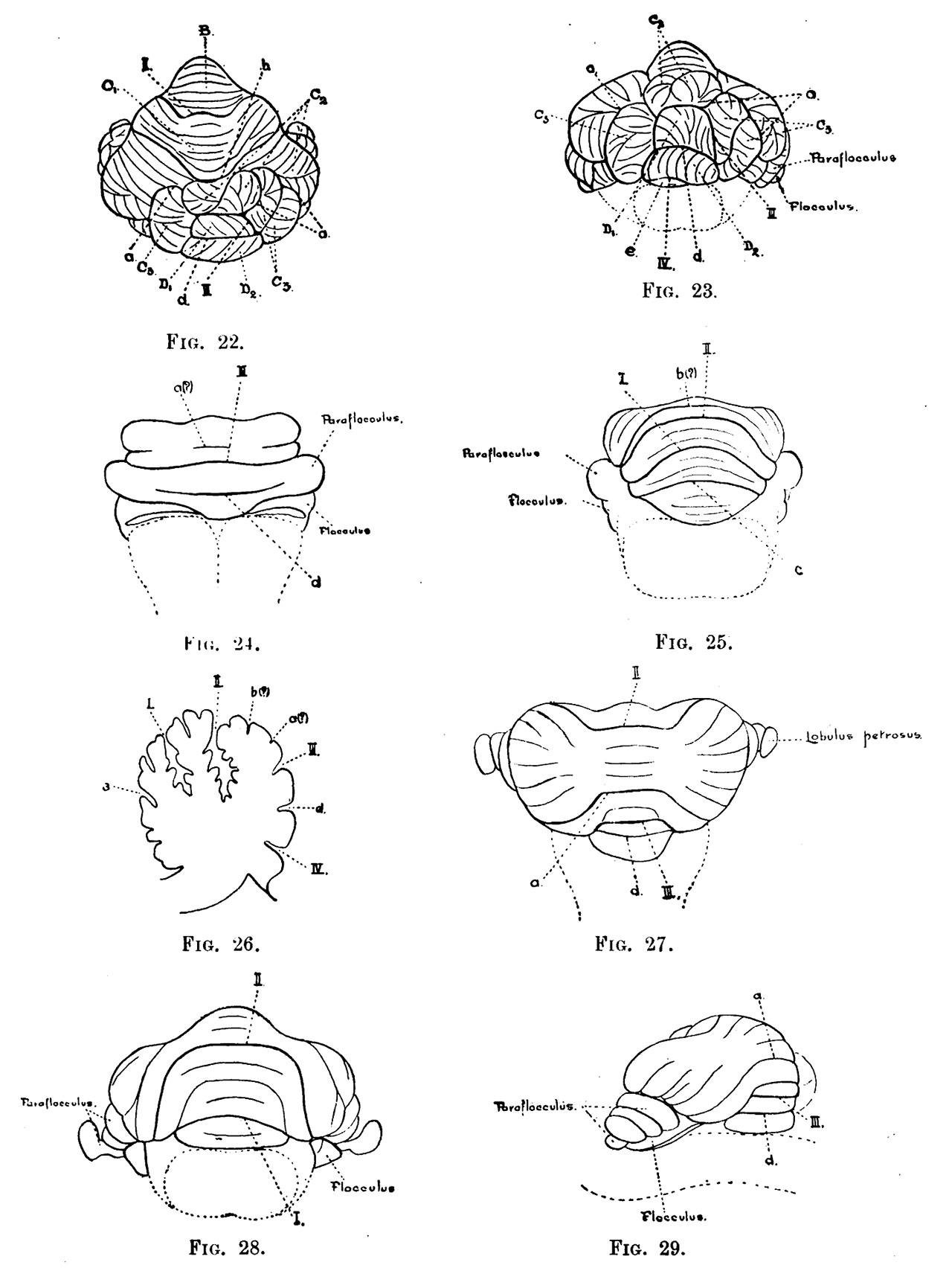
Plate 48
Fig. 22. Bos taurus. Superior view. x 4.
Fig. 23. Bos taurus. Posterior view. x 4.
Fig. 24. horse embryo, 18 weeks old. Posterior view. x 2.
Fig. 25. horse embryo, 18 weeks old. Anterior view.
Fig. 26. horse embryo, 18 weeks old. Mesial sagittal section. The section has been cut slightly obliquely ; the anterior part of the figure therefore shows the fissures, etc., a little more to the right than the posterior part.
Fig. 27. Didelphis azare. Superior view. x 2.
Fig. 28. Didelphis azare. Anterior view. x 2.
Fig. 29. Didelphis azare. Postero-lateral view. x 2.
Fig. 30. Didelphis azare. Mesial sagittal section.
Reference
Bradley OC. The mammalian cerebellum: its lobes and fissures. (1904) J Anat Physiol. 38(4): 448-475. PMID17232617
Cite this page: Hill, M.A. (2024, April 27) Embryology Bradley1904a plate48.jpg. Retrieved from https://embryology.med.unsw.edu.au/embryology/index.php/File:Bradley1904a_plate48.jpg
- © Dr Mark Hill 2024, UNSW Embryology ISBN: 978 0 7334 2609 4 - UNSW CRICOS Provider Code No. 00098G
File history
Click on a date/time to view the file as it appeared at that time.
| Date/Time | Thumbnail | Dimensions | User | Comment | |
|---|---|---|---|---|---|
| current | 18:17, 14 May 2020 | | 1,280 × 1,704 (231 KB) | Z8600021 (talk | contribs) | |
| 17:30, 14 May 2020 | 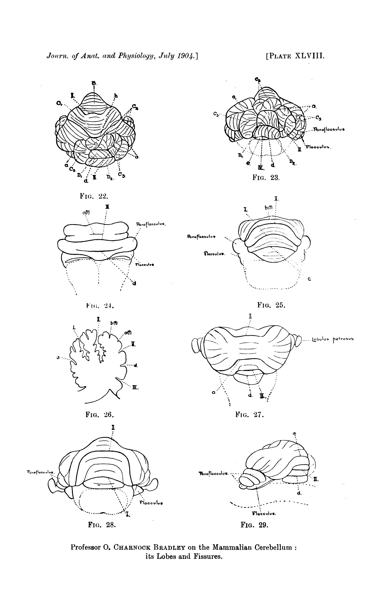 | 1,607 × 2,478 (336 KB) | Z8600021 (talk | contribs) | ==Plate 48== ===Reference=== {{Ref-Bradley1904a}} {{footer}} |
You cannot overwrite this file.
File usage
The following 2 pages use this file: