File:Boyden1932 fig01.jpg

Original file (1,280 × 1,060 pixels, file size: 81 KB, MIME type: image/jpeg)
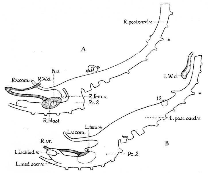
Fig. 1. Graphic reconstruction posterior cardinal veins of a 10 mm human embryo
Graphic reconstruction of right (A) and left (B) posterior cardinal veins of a 10 mm human embryo (no. 134 of Minnesota Collection). X 33.
Drawn from left side, to illustrate greater development of left veins (compare pls. 1 and 2 for orientation). Fus-., area of fusion between left and right blastemas; L. and R.'v.com., vena comitans of left and right umbilical arteries (each vein courses ventrolaterally to its respective umbilical artery until it reaches the caudal umbilical wall. There it anastomoses with the vein of the other side and with the umbilical veins.. These may be the veins which McClure and Butler designate in later stages as the “caudal mesonephric branches of the posterior cardinal veins”); P6..9, second portion of posterior cardinal vein; asterisk, designating level of vitelline artery; 12 and 17, lowest glomeruli of left and right Wolfian bodies, respectively (compare pl. 1).
Reference
Boyden EA. Congenital absence of the kidney - an interpretation based on a 10-mm human embryo exhibiting unilateral renal agenesis. (1932) Anat. Rec. 52(4):325-349.
Cite this page: Hill, M.A. (2024, April 27) Embryology Boyden1932 fig01.jpg. Retrieved from https://embryology.med.unsw.edu.au/embryology/index.php/File:Boyden1932_fig01.jpg
- © Dr Mark Hill 2024, UNSW Embryology ISBN: 978 0 7334 2609 4 - UNSW CRICOS Provider Code No. 00098G
File history
Click on a date/time to view the file as it appeared at that time.
| Date/Time | Thumbnail | Dimensions | User | Comment | |
|---|---|---|---|---|---|
| current | 09:57, 8 September 2017 | | 1,280 × 1,060 (81 KB) | Z8600021 (talk | contribs) | |
| 09:56, 8 September 2017 | 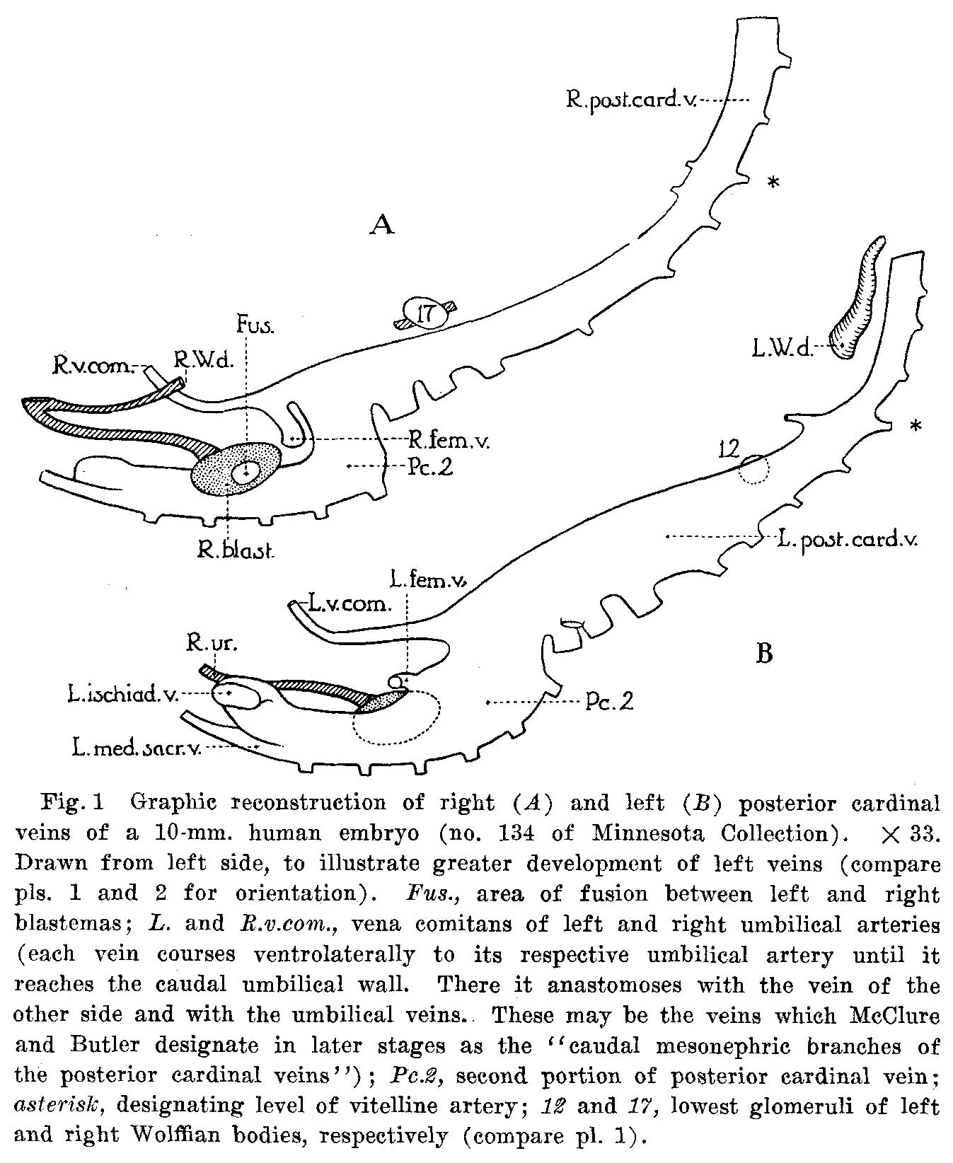 | 1,347 × 1,644 (257 KB) | Z8600021 (talk | contribs) | {{Ref-Boyden1932}} |
You cannot overwrite this file.
File usage
The following page uses this file: