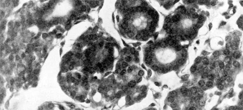
Fig. 10. Section through developing thyroid tissue in 145 mm. human foetus
Colloid in well-established follicles is well shown. The larger spaces lined by endothelium are lymphatic in nature.
Online Editor - Fetus 145mm is about 16 weeks (GA 18 weeks).
| Fertilization and Gestational Age - Crown-Rump Length (ultrasound)
|
Fertilization Age
(days)
|
Gestational Age
GA (week.day)
|
Crown-Rump
Length (mm)
|
| 37
|
5.2
|
1
|
| 38
|
5.3
|
2
|
| 39
|
5.4
|
3
|
| 40
|
55
|
3
|
| 41
|
5.6
|
4
|
| 42 Week 4
|
6
|
4
|
| 43
|
6.1
|
5
|
| 44
|
6.2
|
6
|
| 45
|
6.3
|
7
|
| 46
|
6.4
|
8
|
| 47
|
6.5
|
9
|
| 48
|
6.6
|
10
|
| 49 Week 5
|
7
|
11
|
| 50
|
7.1
|
11
|
| 51
|
7.2
|
12
|
| 52
|
7.3
|
12
|
| 53
|
7.4
|
13
|
| 54
|
7.5
|
14
|
| 55
|
7.6
|
15
|
| 56 Week 6
|
8
|
17
|
| 57
|
8.1
|
18
|
| 58
|
8.2
|
19
|
| 59
|
8.3
|
20
|
| 60
|
8.4
|
21
|
| 61
|
8.5
|
22
|
| 62
|
8.6
|
22
|
| 63 Week 7
|
9
|
23
|
| 64
|
9.1
|
24
|
| 65
|
9.2
|
26
|
| 66
|
9.3
|
27
|
| 67
|
9.4
|
28
|
| 68
|
9.5
|
29
|
| 69
|
9.6
|
31
|
| 70 Week 8
|
10
|
34
|
| 71
|
10.1
|
36
|
| 72
|
10.2
|
37
|
| 73
|
10.3
|
38
|
| 74
|
10.4
|
39
|
| 75
|
10.5
|
39
|
| 76
|
10.6
|
40
|
| 77 Week 9
|
11
|
44
|
| 78
|
11.1
|
45
|
| 79
|
11.2
|
47
|
| 80
|
11.3
|
48
|
| 81
|
11.4
|
52
|
| 82
|
11.5
|
55
|
| 83
|
11.6
|
56
|
| 84 Week 10
|
12
|
57
|
| 85
|
12.1
|
58
|
| 86
|
12.2
|
60
|
| 87
|
12.3
|
61
|
| 88
|
12.4
|
63
|
| 89
|
12.5
|
64
|
| 90
|
12.6
|
65
|
| 91 Week 11
|
13
|
68
|
| 92
|
13.1
|
70
|
| 93
|
13.2
|
72
|
| 94
|
13.3
|
74
|
| 95
|
113.4
|
76
|
| 96
|
135
|
77
|
| 97
|
13.6
|
80
|
| 98 Week 12
|
14
|
81
|
| 99
|
14.1
|
84
|
| 100
|
14.2
|
85
|
| 101
|
14.3
|
86
|
| 102
|
14.4
|
87
|
Reference: Table data measured by ultrasound, adapted from Westerway (2015) PDF and[1]
- Links: ultrasound | Fetal Development
|
|
| Historic Disclaimer - information about historic embryology pages
|
| Pages where the terms "Historic" (textbooks, papers, people, recommendations) appear on this site, and sections within pages where this disclaimer appears, indicate that the content and scientific understanding are specific to the time of publication. This means that while some scientific descriptions are still accurate, the terminology and interpretation of the developmental mechanisms reflect the understanding at the time of original publication and those of the preceding periods, these terms, interpretations and recommendations may not reflect our current scientific understanding. (More? Embryology History | Historic Embryology Papers)
|
- Links: fig 1 | fig 2 | fig 3a | fig 3b | fig 4 | fig 5 | fig 6 | fig 7 | fig 8 | fig 9 | fig 10 | 1950 Boyd | Thyroid | Parathyroid | Thymus | Historic Papers
Reference
Boyd JD. Development of the thyroid and parathyroid glands and the thymus. (1950) Ann R Coll Surg Engl. 7(6): 455-71. PMID 14790564
Cite this page: Hill, M.A. (2024, May 6) Embryology Boyd1950 fig10.jpg. Retrieved from https://embryology.med.unsw.edu.au/embryology/index.php/File:Boyd1950_fig10.jpg
- What Links Here?
- © Dr Mark Hill 2024, UNSW Embryology ISBN: 978 0 7334 2609 4 - UNSW CRICOS Provider Code No. 00098G
- ↑ Westerway SC, Davison A & Cowell S. (2000). Ultrasonic fetal measurements: new Australian standards for the new millennium. Aust N Z J Obstet Gynaecol , 40, 297-302. PMID: 11065037
File history
Click on a date/time to view the file as it appeared at that time.
| Date/Time | Thumbnail | Dimensions | User | Comment |
|---|
| current | 08:11, 21 March 2017 |  | 1,000 × 453 (114 KB) | Z8600021 (talk | contribs) | |
| 08:07, 21 March 2017 |  | 1,336 × 779 (295 KB) | Z8600021 (talk | contribs) | |
You cannot overwrite this file.
File usage
The following page uses this file:
This file contains additional information, probably added from the digital camera or scanner used to create or digitise it.
If the file has been modified from its original state, some details may not fully reflect the modified file.


