File:Boyd1950 fig04.jpg
From Embryology

Size of this preview: 682 × 599 pixels.
Original file (800 × 703 pixels, file size: 102 KB, MIME type: image/jpeg)
Fig. 4. Section of thymus of 140 mm human foetus
To show three Hassall‘s corpuscles in the medulla. ( X 1000)
| Historic Disclaimer - information about historic embryology pages |
|---|
 |
- Links: fig 1 | fig 2 | fig 3a | fig 3b | fig 4 | fig 5 | fig 6 | fig 7 | fig 8 | fig 9 | fig 10 | 1950 Boyd | Thyroid | Parathyroid | Thymus | Historic Papers
Reference
Boyd JD. Development of the thyroid and parathyroid glands and the thymus. (1950) Ann R Coll Surg Engl. 7(6): 455-71. PMID 14790564
Cite this page: Hill, M.A. (2024, April 27) Embryology Boyd1950 fig04.jpg. Retrieved from https://embryology.med.unsw.edu.au/embryology/index.php/File:Boyd1950_fig04.jpg
- © Dr Mark Hill 2024, UNSW Embryology ISBN: 978 0 7334 2609 4 - UNSW CRICOS Provider Code No. 00098G
File history
Click on a date/time to view the file as it appeared at that time.
| Date/Time | Thumbnail | Dimensions | User | Comment | |
|---|---|---|---|---|---|
| current | 07:14, 21 March 2017 | | 800 × 703 (102 KB) | Z8600021 (talk | contribs) | |
| 07:13, 21 March 2017 | 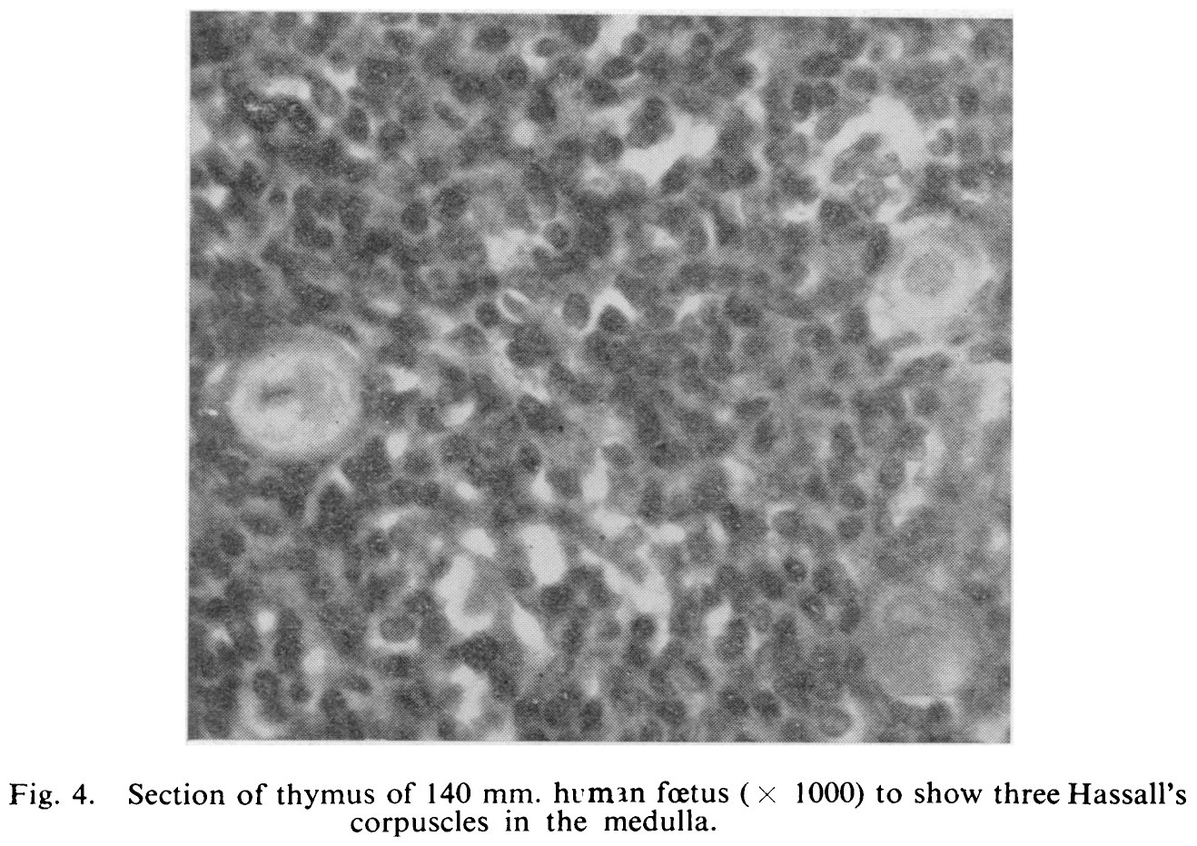 | 1,318 × 932 (269 KB) | Z8600021 (talk | contribs) |
You cannot overwrite this file.
File usage
The following page uses this file: