File:Bonnot1906 plate04.jpg
Original file (1,161 × 1,675 pixels, file size: 122 KB, MIME type: image/jpeg)
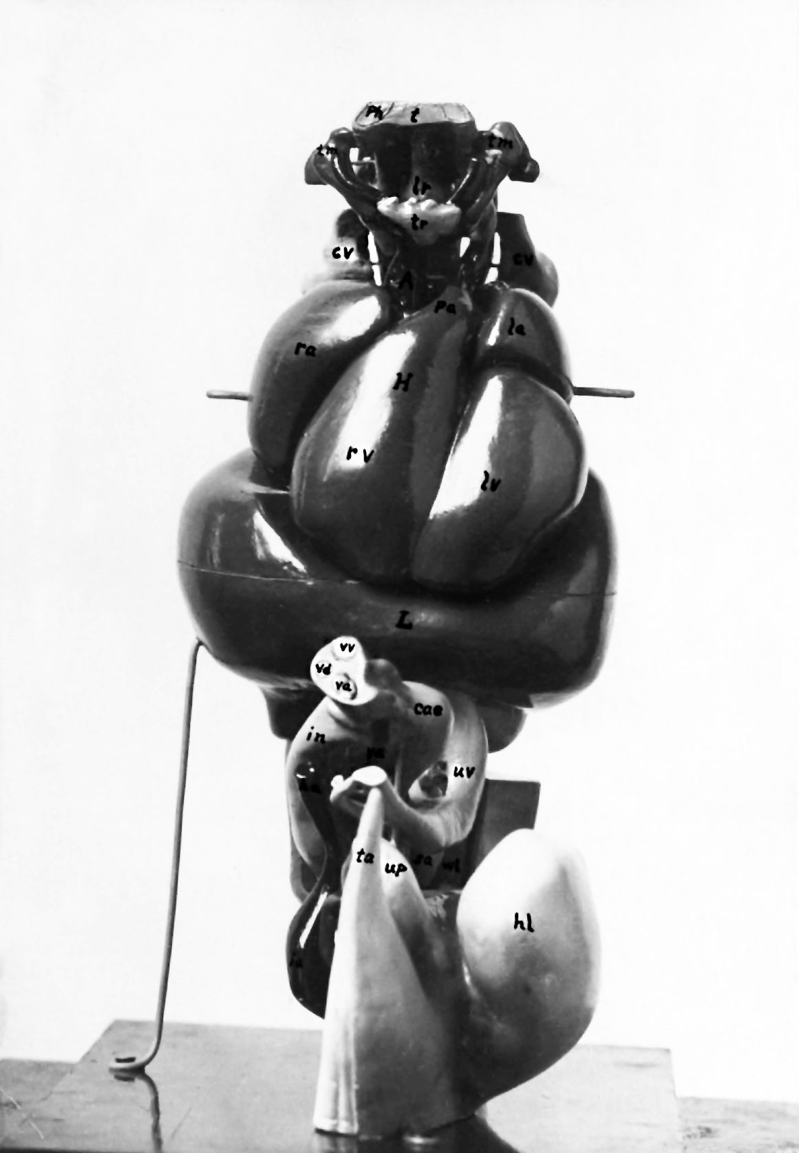
Plate 4. Photograph of the anterior view of the model
The small uper part shows the pharynx and its appendages, and the branchial arteries. The large middle part of the model represents the heart above and the liver below. Below the liver are the intestines, yolk stalk and umbilical vessels.
A, ascending aorta; cae, caocum; cv, anterior cardinal veins; H, heart; ha, hypogastric artery; hl, hind
limb; ia. iliac artery; in, intestine; L, liver la, left auricle; lr, Larynx; lv, left ventricle; pa, pulmonary artery; ph, pharynx; rs, right auriole; rv, rimht
ventricle; ea, sexual anlsge; t, tongue; ta, tail; tm,
thyxus; tr, thyroid gland; up, urinogenital papilla; uv, umbilical vein; va, vitelline artery; vd, vétclline duet; vv, vitelline vein; wl, Wolffian bodies.
(From Bonnet's thesis)
Reference
Bonnet E. and Severs R. On the structure of a human embryo eleven millimeters in length. (1906) Anat. Anz., 29: 452-459.
Cite this page: Hill, M.A. (2024, April 28) Embryology Bonnot1906 plate04.jpg. Retrieved from https://embryology.med.unsw.edu.au/embryology/index.php/File:Bonnot1906_plate04.jpg
- © Dr Mark Hill 2024, UNSW Embryology ISBN: 978 0 7334 2609 4 - UNSW CRICOS Provider Code No. 00098G
File history
Click on a date/time to view the file as it appeared at that time.
| Date/Time | Thumbnail | Dimensions | User | Comment | |
|---|---|---|---|---|---|
| current | 17:06, 19 December 2016 | | 1,161 × 1,675 (122 KB) | Z8600021 (talk | contribs) |
You cannot overwrite this file.
File usage
The following 2 pages use this file: