File:Bonnot1906 fig03.jpg: Difference between revisions
(===Reference=== {{Ref-Bonnot1906}} {{Footer}} Category:Carnegie Stage 16Category:Week 6 Category:1900's) |
mNo edit summary |
||
| Line 1: | Line 1: | ||
==Fig. 3. From a photograph of the right view of the model== | |||
On this side, the lower part of the body wall is not seen as on the left side, but has been dissected out to the mid-sagittal plane, giving a side view of the spinal cord, notochord, the descending aorta with its branches, and the pelvic viscera. | |||
Explanation (in addition to letters shown in [[:File:Bonnot1906 fig02.jpg|Fig. 2]]): a‘ right aortic arch. al allantois. b Wolffian duet. ca caudal artery. cl cloaca. d duodenum. ha hypogastrie artery. i intestine. n notochord. o gall bladder. r rectum. ra right auricle. rv right ventricle. sc spinal cord. sv sinus venosus. u ureter. Its T-shaped upper extremity, forming the anlage of the permanent kidney, is almost entirely hidden by the hypogastric artery. up urinogenital papilla. vv vitelline (omphalomesenteric) vein. | |||
===Reference=== | ===Reference=== | ||
Revision as of 16:47, 19 December 2016
Fig. 3. From a photograph of the right view of the model
On this side, the lower part of the body wall is not seen as on the left side, but has been dissected out to the mid-sagittal plane, giving a side view of the spinal cord, notochord, the descending aorta with its branches, and the pelvic viscera.
Explanation (in addition to letters shown in Fig. 2): a‘ right aortic arch. al allantois. b Wolffian duet. ca caudal artery. cl cloaca. d duodenum. ha hypogastrie artery. i intestine. n notochord. o gall bladder. r rectum. ra right auricle. rv right ventricle. sc spinal cord. sv sinus venosus. u ureter. Its T-shaped upper extremity, forming the anlage of the permanent kidney, is almost entirely hidden by the hypogastric artery. up urinogenital papilla. vv vitelline (omphalomesenteric) vein.
Reference
Bonnet E. and Severs R. On the structure of a human embryo eleven millimeters in length. (1906) Anat. Anz., 29: 452-459.
Cite this page: Hill, M.A. (2024, April 26) Embryology Bonnot1906 fig03.jpg. Retrieved from https://embryology.med.unsw.edu.au/embryology/index.php/File:Bonnot1906_fig03.jpg
- © Dr Mark Hill 2024, UNSW Embryology ISBN: 978 0 7334 2609 4 - UNSW CRICOS Provider Code No. 00098G
File history
Click on a date/time to view the file as it appeared at that time.
| Date/Time | Thumbnail | Dimensions | User | Comment | |
|---|---|---|---|---|---|
| current | 16:44, 19 December 2016 | 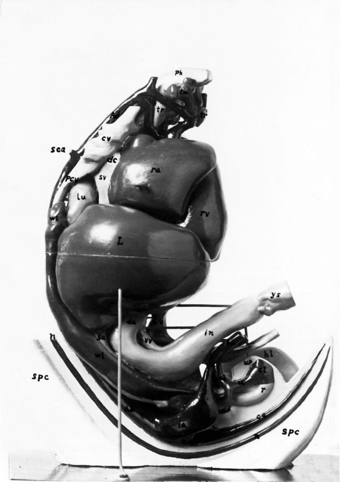 | 1,192 × 1,689 (154 KB) | Z8600021 (talk | contribs) | ===Reference=== {{Ref-Bonnot1906}} {{Footer}} Category:Carnegie Stage 16Category:Week 6 Category:1900's |
You cannot overwrite this file.
File usage
The following 2 pages use this file: