File:Blood Smear Slide 02.jpg: Difference between revisions
From Embryology
mNo edit summary |
mNo edit summary |
||
| Line 6: | Line 6: | ||
:'''Links:''' [[ANAT2241 Blood|Science - Blood Histology]] | [[Histology]] | [[Cardiovascular System - Blood Development|Blood Development]] | :'''Links:''' [[:File:Blood Smear Slide 01.jpg|image - slide 1]] | [[:File:Blood Smear Slide 02.jpg|image - slide 2]] | [[ANAT2241 Blood|Science - Blood Histology]] | [[Histology]] | [[Cardiovascular System - Blood Development|Blood Development]] | ||
===Reference=== | ===Reference=== | ||
CDC/ Steven Glenn, Laboratory & Consultation Division (1979) | CDC/ Steven Glenn, Laboratory & Consultation Division (1979) | ||
Latest revision as of 10:04, 13 June 2015
Blood Smear Slide
This prepared slide offers an example of the appearance of “marginal quality” thick and thin film blood smears.
Though they are more difficult to read, thick blood films are more sensitive in detecting malaria parasites because the blood is concentrated, allowing a greater volume of blood to be examined.
- Links: image - slide 1 | image - slide 2 | Science - Blood Histology | Histology | Blood Development
Reference
CDC/ Steven Glenn, Laboratory & Consultation Division (1979)
Copyright
This image is in the public domain and thus free of any copyright restrictions. As a matter of courtesy we request that the content provider be credited and notified in any public or private usage of this image.
5894_lores.jpg
File history
Click on a date/time to view the file as it appeared at that time.
| Date/Time | Thumbnail | Dimensions | User | Comment | |
|---|---|---|---|---|---|
| current | 09:55, 13 June 2015 | 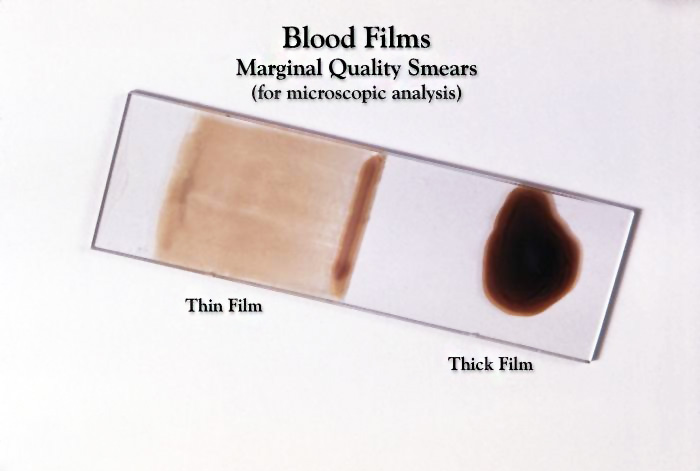 | 700 × 471 (47 KB) | Z8600021 (talk | contribs) |
You cannot overwrite this file.
File usage
The following page uses this file: