File:Blood-thymus barrier EM01.jpg: Difference between revisions
mNo edit summary |
mNo edit summary |
||
| Line 1: | Line 1: | ||
==Blood-Thymus Barrier - Electron micrograph== | ==Blood-Thymus Barrier - Electron micrograph== | ||
Capillary of the | Capillary of the {{mouse}} {{thymus}} cortex. X 27,000. | ||
In this specimen care has been taken to retain the blood in the vascular bed during fixation. The capillary, lumen is wide open and contains both plasma and red blood cells; in the cytoplasm of the attenuated endo- thelial cells (End), plasmalemmal vesicles are few in numbers. | In this specimen care has been taken to retain the blood in the vascular bed during fixation. The capillary, lumen is wide open and contains both plasma and red blood cells; in the cytoplasm of the attenuated endo- thelial cells (End), plasmalemmal vesicles are few in numbers. | ||
| Line 8: | Line 8: | ||
Below and on the left, the parenchymal front bounding the vascular adventia is represented by a process of a reticular cell. | Below and on the left, the parenchymal front bounding the vascular adventia is represented by a process of a reticular cell. | ||
===Reference=== | ===Reference=== | ||
Revision as of 16:51, 10 May 2018
Blood-Thymus Barrier - Electron micrograph
Capillary of the mouse thymus cortex. X 27,000.
In this specimen care has been taken to retain the blood in the vascular bed during fixation. The capillary, lumen is wide open and contains both plasma and red blood cells; in the cytoplasm of the attenuated endo- thelial cells (End), plasmalemmal vesicles are few in numbers.
The vessel is enclosed in a coaxial canal formed by the surrounding thymic parenchymal cells; a macrophage containing residual bodies (asterisks) encompasses most of the circumference of the vessel and sends processes into the vascular adventitia (Adv).
Below and on the left, the parenchymal front bounding the vascular adventia is represented by a process of a reticular cell.
Reference
Raviola E & Karnovsky MJ. (1972). Evidence for a blood-thymus barrier using electron-opaque tracers. J. Exp. Med. , 136, 466-98. PMID: 4115129
Fig. 2. modified in size, contract and labelling.
Copyright
Rockefeller University Press - Copyright Policy This article is distributed under the terms of an Attribution–Noncommercial–Share Alike–No Mirror Sites license for the first six months after the publication date (see http://www.jcb.org/misc/terms.shtml). After six months it is available under a Creative Commons License (Attribution–Noncommercial–Share Alike 4.0 Unported license, as described at https://creativecommons.org/licenses/by-nc-sa/4.0/ ). (More? Help:Copyright Tutorial)
File history
Click on a date/time to view the file as it appeared at that time.
| Date/Time | Thumbnail | Dimensions | User | Comment | |
|---|---|---|---|---|---|
| current | 13:54, 10 May 2018 | 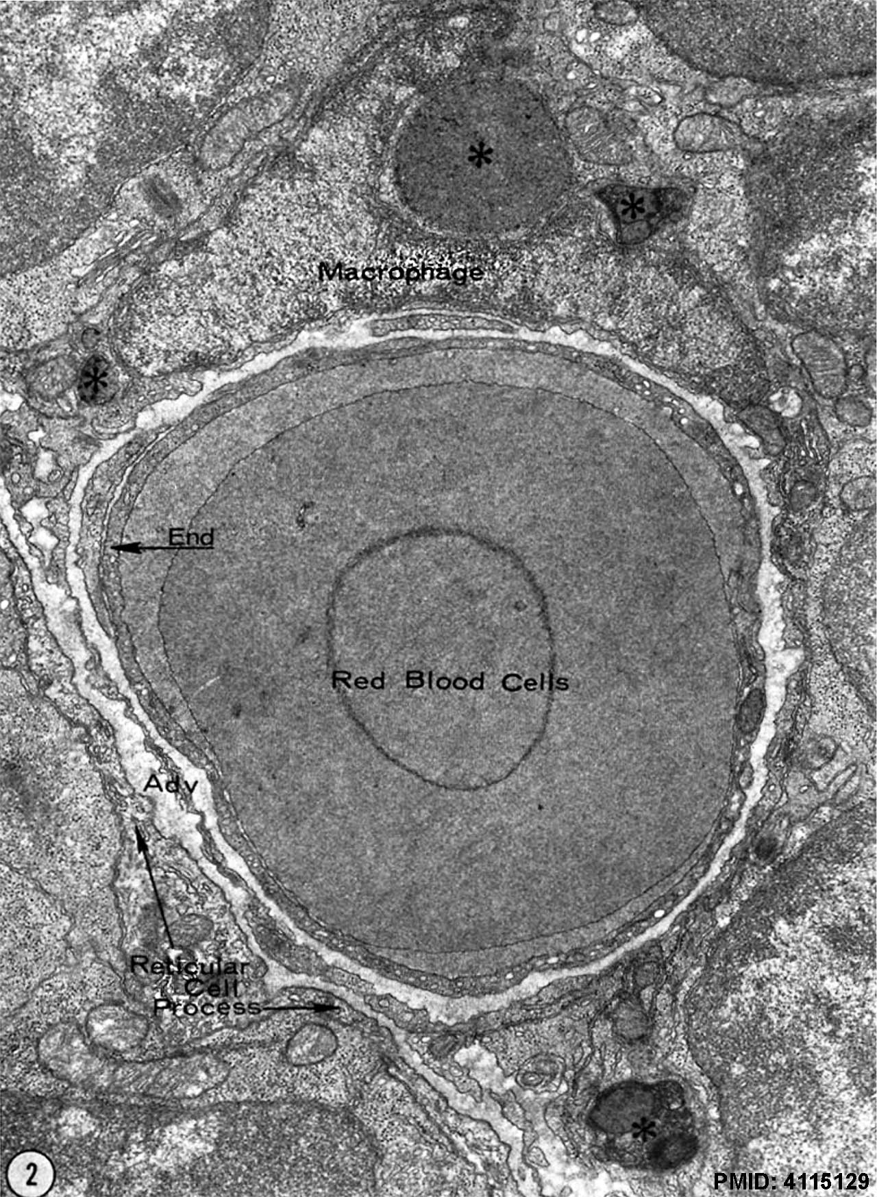 | 1,280 × 1,747 (375 KB) | Z8600021 (talk | contribs) | |
| 13:54, 10 May 2018 | 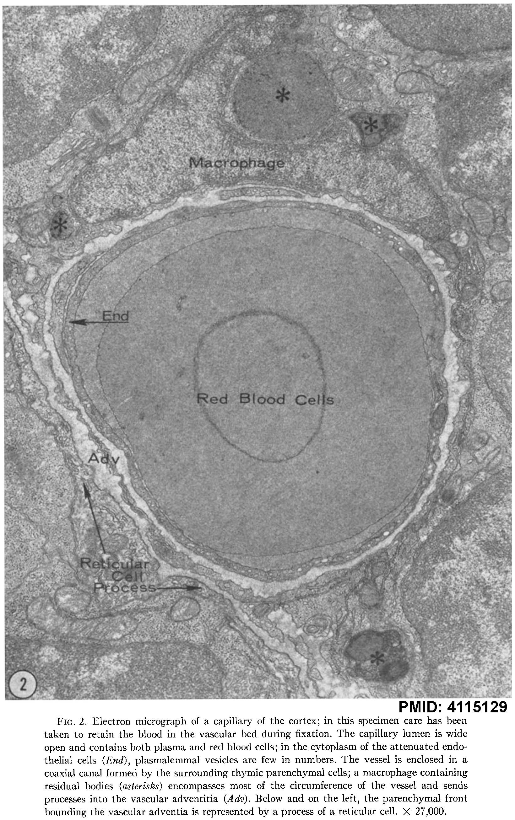 | 1,640 × 2,613 (478 KB) | Z8600021 (talk | contribs) | {{#pmid:4115129}} |
You cannot overwrite this file.
File usage
The following 2 pages use this file: