File:Blastomere isolation.jpg
Original file (1,200 × 961 pixels, file size: 95 KB, MIME type: image/jpeg)
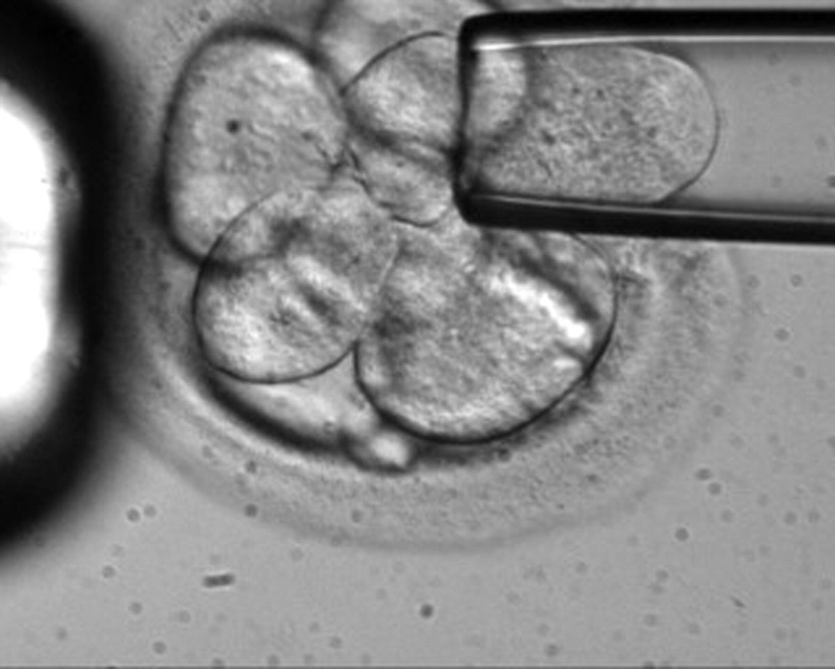
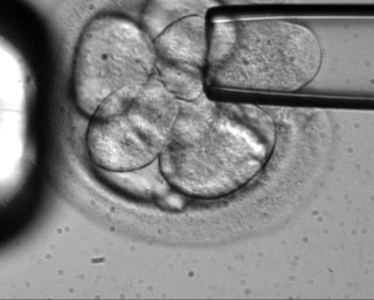
Blastomere Isolation
This is early morula stage still enclosed by the zona pellucida.
- A single blastomere cell is being drawn into the pipette (right) from the solid cellular mass.
- A second larger pipette (left) holds the morula in place.
One of the new methods for generating embryonic stem cells (ESCs) involves using a single blastomere.
Reference
Leslie M. (2006). The action behind the words: embryonic stem cell research marches on. J. Cell Biol. , 174, 743-6. PMID: 16966416 DOI.
Copyright
Rockefeller University Press - Copyright Policy This article is distributed under the terms of an Attribution–Noncommercial–Share Alike–No Mirror Sites license for the first six months after the publication date (see http://www.jcb.org/misc/terms.shtml). After six months it is available under a Creative Commons License (Attribution–Noncommercial–Share Alike 4.0 Unported license, as described at https://creativecommons.org/licenses/by-nc-sa/4.0/ ). (More? Help:Copyright Tutorial)
Original file name: Figure 1. http://jcb.rupress.org/content/174/6/743/F1.expansion.html
Cite this page: Hill, M.A. (2024, April 27) Embryology Blastomere isolation.jpg. Retrieved from https://embryology.med.unsw.edu.au/embryology/index.php/File:Blastomere_isolation.jpg
- © Dr Mark Hill 2024, UNSW Embryology ISBN: 978 0 7334 2609 4 - UNSW CRICOS Provider Code No. 00098G
File history
Click on a date/time to view the file as it appeared at that time.
| Date/Time | Thumbnail | Dimensions | User | Comment | |
|---|---|---|---|---|---|
| current | 23:52, 24 May 2011 | | 1,200 × 961 (95 KB) | S8600021 (talk | contribs) | One of the new methods for generating embryonic stem cells (ESCs) involves using a single blastomere. Original file name: Figure 1. http://jcb.rupress.org/content/174/6/743/F1.expansion.html ==Reference== <pubmed>16966416</pubmed> {{JCB}} |
You cannot overwrite this file.
File usage
The following page uses this file: