File:Berkeley1905 fig03.jpg

Original file (1,000 × 966 pixels, file size: 158 KB, MIME type: image/jpeg)
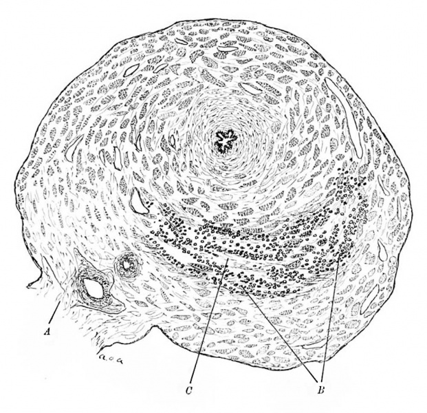
Fig. 3 illustrates a section taken about a millimetre nearer the centre of the gestation
Evidence of foetal invasion is here very marked along crescentic delaminations of the muscle wall on the side of attachment to the mesosalpinx. Along these delamintations the troplioblast cells appear to be proceeding in such a way that the walls of the spaces are very deeply infiltrated by them. The maternal tisue in their vicinity is broken up, and is undergoing a fibrinous change. There is little or no evidence of proliferation of the maternal cells or of an active reply of any kind by them to the destructive action of the trophoblast, whose cell are mainly of two kinds—large mononuclear cells and irregular multinuclear syncytia. Here and there are seen multinuclear cells, which are probably to be regarded as intermediate stages between these.
Reference
Berkeley C. and Bonney V. Tubal Gestation - A Pathological Study. (1905) Brit. J. Obst. and Gyn. 7(2): 78-96.
Cite this page: Hill, M.A. (2024, April 30) Embryology Berkeley1905 fig03.jpg. Retrieved from https://embryology.med.unsw.edu.au/embryology/index.php/File:Berkeley1905_fig03.jpg
- © Dr Mark Hill 2024, UNSW Embryology ISBN: 978 0 7334 2609 4 - UNSW CRICOS Provider Code No. 00098G
File history
Click on a date/time to view the file as it appeared at that time.
| Date/Time | Thumbnail | Dimensions | User | Comment | |
|---|---|---|---|---|---|
| current | 10:40, 5 November 2017 | | 1,000 × 966 (158 KB) | Z8600021 (talk | contribs) | |
| 10:39, 5 November 2017 | 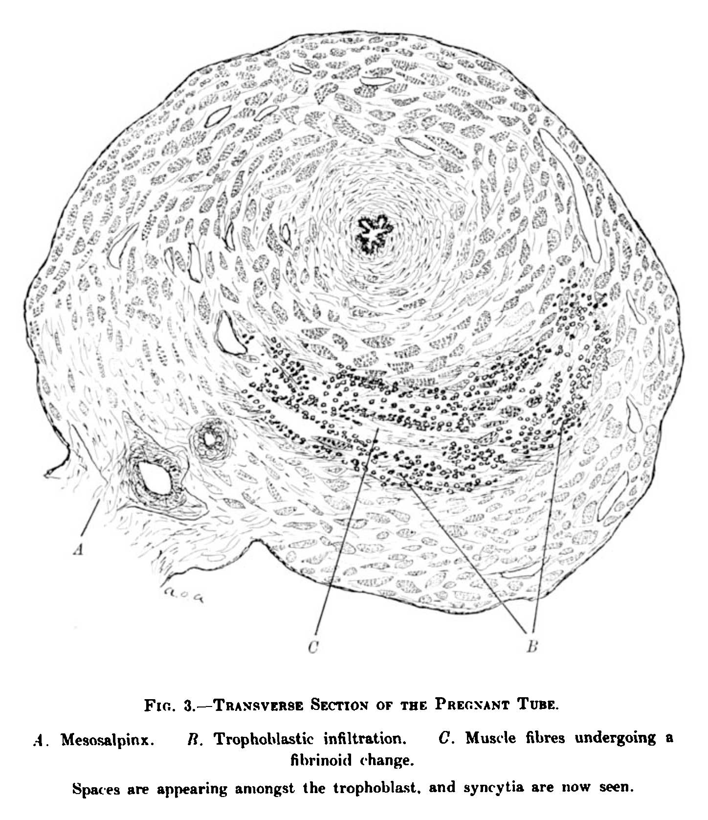 | 1,448 × 1,676 (249 KB) | Z8600021 (talk | contribs) |
You cannot overwrite this file.
File usage
The following page uses this file: