File:Berkeley1905 fig01.jpg

Original file (1,000 × 635 pixels, file size: 86 KB, MIME type: image/jpeg)
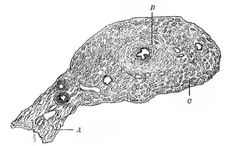
Fig. 1. Transverse section of the pregnant tube between the gestational sac and the uterine cornu
A. Mesosalpinx. B. Circular Muscle Coat. C. Longitudinal Muscle Coat.
Transverse section of the tube on the abdominal side of the gestation sac a illustrates a transverse section of the tube on the abdominal side of the gestation sac and presents nothing abnormal.
It will be seen from the simple arrangement of the plicee and the relative thickness of the muscle that it is taken from the isthmic portion of the tube. An attentive examination of the section reveals no trace of foetal cells, either in the tissues or in the vessels.
Reference
Berkeley C. and Bonney V. Tubal Gestation - A Pathological Study. (1905) Brit. J. Obst. and Gyn. 7(2): 78-96.
Cite this page: Hill, M.A. (2024, May 21) Embryology Berkeley1905 fig01.jpg. Retrieved from https://embryology.med.unsw.edu.au/embryology/index.php/File:Berkeley1905_fig01.jpg
- © Dr Mark Hill 2024, UNSW Embryology ISBN: 978 0 7334 2609 4 - UNSW CRICOS Provider Code No. 00098G
File history
Click on a date/time to view the file as it appeared at that time.
| Date/Time | Thumbnail | Dimensions | User | Comment | |
|---|---|---|---|---|---|
| current | 10:34, 5 November 2017 | | 1,000 × 635 (86 KB) | Z8600021 (talk | contribs) | |
| 10:31, 5 November 2017 | 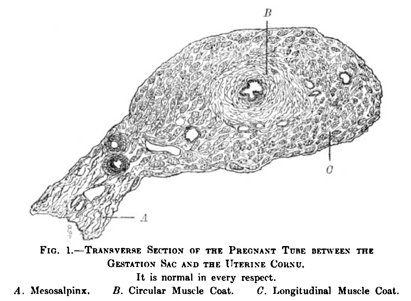 | 1,329 × 975 (122 KB) | Z8600021 (talk | contribs) | ===Reference=== {{Ref-Berkeley1905}} |
You cannot overwrite this file.
File usage
The following page uses this file: