File:Baumgartner1916 plate01.jpg

Original file (1,692 × 2,228 pixels, file size: 308 KB, MIME type: image/jpeg)
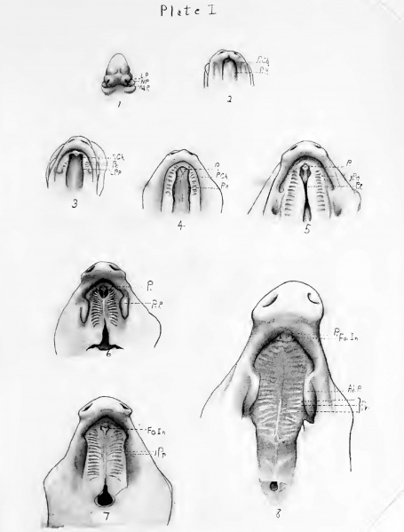
Plate 1. Pig Palate Development
Fig. 1. Frontal view of the head of the 12 mm. pig embryo.
Fig. 2. Ventral view of the roof of the primitive mouth of the 17 mm. embryo.
Fig. 3. Ventral view of the roof of the primitive mouth of the 20 mm. embryo.
Fig.4. Ventral view of the roof of the primitive mouth of the 25 mm. embryo.
Fig. 5. Ventral View of the roof of the primitive mouth of the 27 mm. embryo.
Fig. 6. Ventral view of the roof of the secondary mouth of the 50 mm. embryo.
Fig. 7. Ventral view of the roof of the secondary mouth of the 39 mm. embryo.
Fig. 8. Ventral View of the roof of the secondary mouth of the 70 mm. embryo.
- Links: plate 1 | plate 2 | Palate Development | Pig Development
Reference
Baumgartner RA. Development of the palate and the definitive choanae in the pig. (1916) Thesis, University of Illinois.
Cite this page: Hill, M.A. (2024, April 27) Embryology Baumgartner1916 plate01.jpg. Retrieved from https://embryology.med.unsw.edu.au/embryology/index.php/File:Baumgartner1916_plate01.jpg
- © Dr Mark Hill 2024, UNSW Embryology ISBN: 978 0 7334 2609 4 - UNSW CRICOS Provider Code No. 00098G
File history
Click on a date/time to view the file as it appeared at that time.
| Date/Time | Thumbnail | Dimensions | User | Comment | |
|---|---|---|---|---|---|
| current | 10:13, 11 June 2016 | | 1,692 × 2,228 (308 KB) | Z8600021 (talk | contribs) | ==Pig Palate Development== ===Reference=== Baumgartner RA. Development of the palate and the definitive choanae in the pig (1916) Thesis University of Illinois. {{Footer}} |
You cannot overwrite this file.
File usage
The following page uses this file: