File:Bast1933 fig33.jpg

Original file (1,000 × 673 pixels, file size: 73 KB, MIME type: image/jpeg)
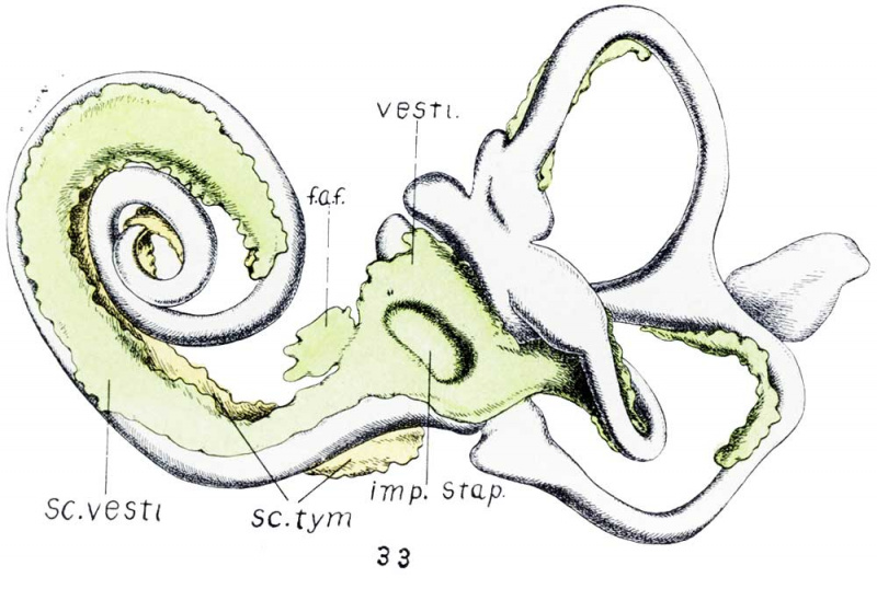
Figs. 33 Human Fetus CRL 50 mm
Fig. 33. Fetus 86; size, 50 mm. crown-rump length; age, about 10 weeks. Anterior view of a model of the otic labyrinth showing the development of the periotic tissue and spaces and the fissula ante fenestram.
In figures 33 and 34, vesti. indicates vestibule; f.a.f., fissula ante fenestram; S c. 'vesti., scala vestibuli; Sc. tym., scala tympani; imp. Sta.p., impression of stapes; Sac. end., endolyrnphatic duct; Sm:., sacculus; Ut1'z'c., utriculus; Duct. coch-., cochlear duct.
Online Editor - CRL 50 mm about Week 10 development.
Reference
Bast TH. Development of the otic capsule II. The origin, development and significance of the fissula ante fenestram and its relation to otosclerotic foci. (1933) Arch. Otolaryng. 18(1):
Cite this page: Hill, M.A. (2024, April 28) Embryology Bast1933 fig33.jpg. Retrieved from https://embryology.med.unsw.edu.au/embryology/index.php/File:Bast1933_fig33.jpg
- © Dr Mark Hill 2024, UNSW Embryology ISBN: 978 0 7334 2609 4 - UNSW CRICOS Provider Code No. 00098G
File history
Click on a date/time to view the file as it appeared at that time.
| Date/Time | Thumbnail | Dimensions | User | Comment | |
|---|---|---|---|---|---|
| current | 16:04, 22 October 2017 | | 1,000 × 673 (73 KB) | Z8600021 (talk | contribs) |
You cannot overwrite this file.
File usage
The following page uses this file: