File:Bast1931 plate01.jpg: Difference between revisions
mNo edit summary |
mNo edit summary |
||
| Line 14: | Line 14: | ||
{{Footer}} | {{Footer}} | ||
[[Category:Hearing]][[Category:Fetus]] | |||
Revision as of 17:26, 4 October 2017
Plate 1
This is a drawing of part of a model of the internal ear of a 150-mm. (C.R.) human fetus, age about eighteen and one-half weeks. It is drawn to show the arterial blood supply to the otic capsule.
The periotic labyrinth is shown in yellow, the ossified portion of the capsule in black, and the arteries in red.
The numbers 1, 2, 3, 4, 8, and 10 represent the approximate point of origin of the corresponding ossification centers (Bast, ’30).
The part of the capsule which is still cartilage is not drawn.
Reference
Bast TH. Blood supply of the otic capsule of a 150 mm (C.R.) human fetus. (1931) Anat. Rec. 48: 141-151.
Cite this page: Hill, M.A. (2024, April 27) Embryology Bast1931 plate01.jpg. Retrieved from https://embryology.med.unsw.edu.au/embryology/index.php/File:Bast1931_plate01.jpg
- © Dr Mark Hill 2024, UNSW Embryology ISBN: 978 0 7334 2609 4 - UNSW CRICOS Provider Code No. 00098G
File history
Click on a date/time to view the file as it appeared at that time.
| Date/Time | Thumbnail | Dimensions | User | Comment | |
|---|---|---|---|---|---|
| current | 17:24, 4 October 2017 | 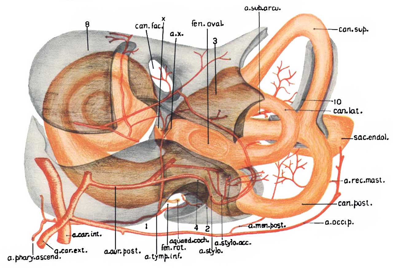 | 1,280 × 871 (113 KB) | Z8600021 (talk | contribs) | |
| 17:24, 4 October 2017 | 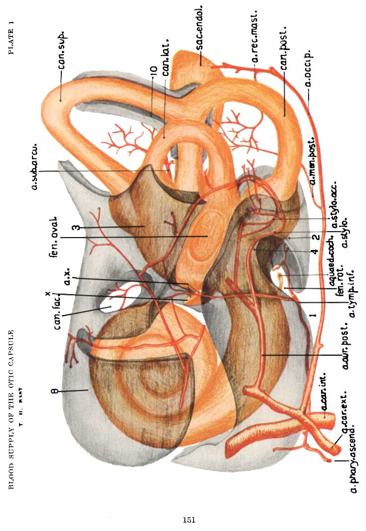 | 1,474 × 2,140 (228 KB) | Z8600021 (talk | contribs) | ===Reference=== {{Ref-Bast1931}} |
You cannot overwrite this file.
File usage
The following page uses this file: