File:Bartelmez1923 fig02.jpg

Original file (1,295 × 2,189 pixels, file size: 218 KB, MIME type: image/jpeg)
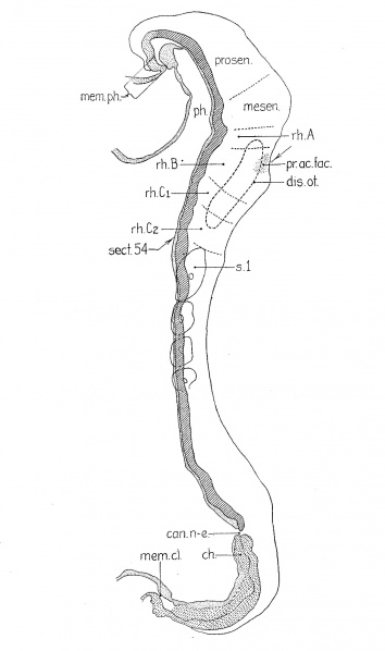
Fig. 2 A reconstruction of a four—somite embryo
(H279, U. of C. Coll.); magnified 200 diameters and reduced one half in reproduction. The plane of section is indicated by the position of section 54 which was shown as figure 1 in my 1922 paper. camn-e., neurenteric canal; ch., chorda; disc.o£., otic disc; mom. cl., cloacal membrane (the entoderm has shrunken away from the cctoderm) ; mem. ph., pharyngeal membrane; mesen., midbrain; ph., pharynx; pr.ac.fac., acoustic0-facial primordium; prosen., forebrain; rh.A, rh.B, first and second hindbrain segments; rh..C'1 and 7'h.C'2 have resulted from the division of rh.C’ in the previous embryo; rh.B is identical with the definitive fourth rhombomere, rh.C'; with the fifth.
File history
Click on a date/time to view the file as it appeared at that time.
| Date/Time | Thumbnail | Dimensions | User | Comment | |
|---|---|---|---|---|---|
| current | 22:40, 7 June 2016 |  | 1,295 × 2,189 (218 KB) | Z8600021 (talk | contribs) | ==Fig. 2 A reconstruction of a four—somite embryo== (H279, U. of C. Coll.); magnified 200 diameters and reduced one half in reproduction. The plane of section is indicated by the position of section 54 which was shown as figure 1 in my 1922 paper. c... |
You cannot overwrite this file.
File usage
The following 2 pages use this file: