File:Bardeen1908a fig01.jpg
Original file (1,000 × 1,373 pixels, file size: 76 KB, MIME type: image/jpeg)
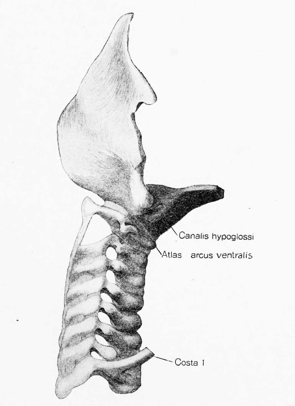
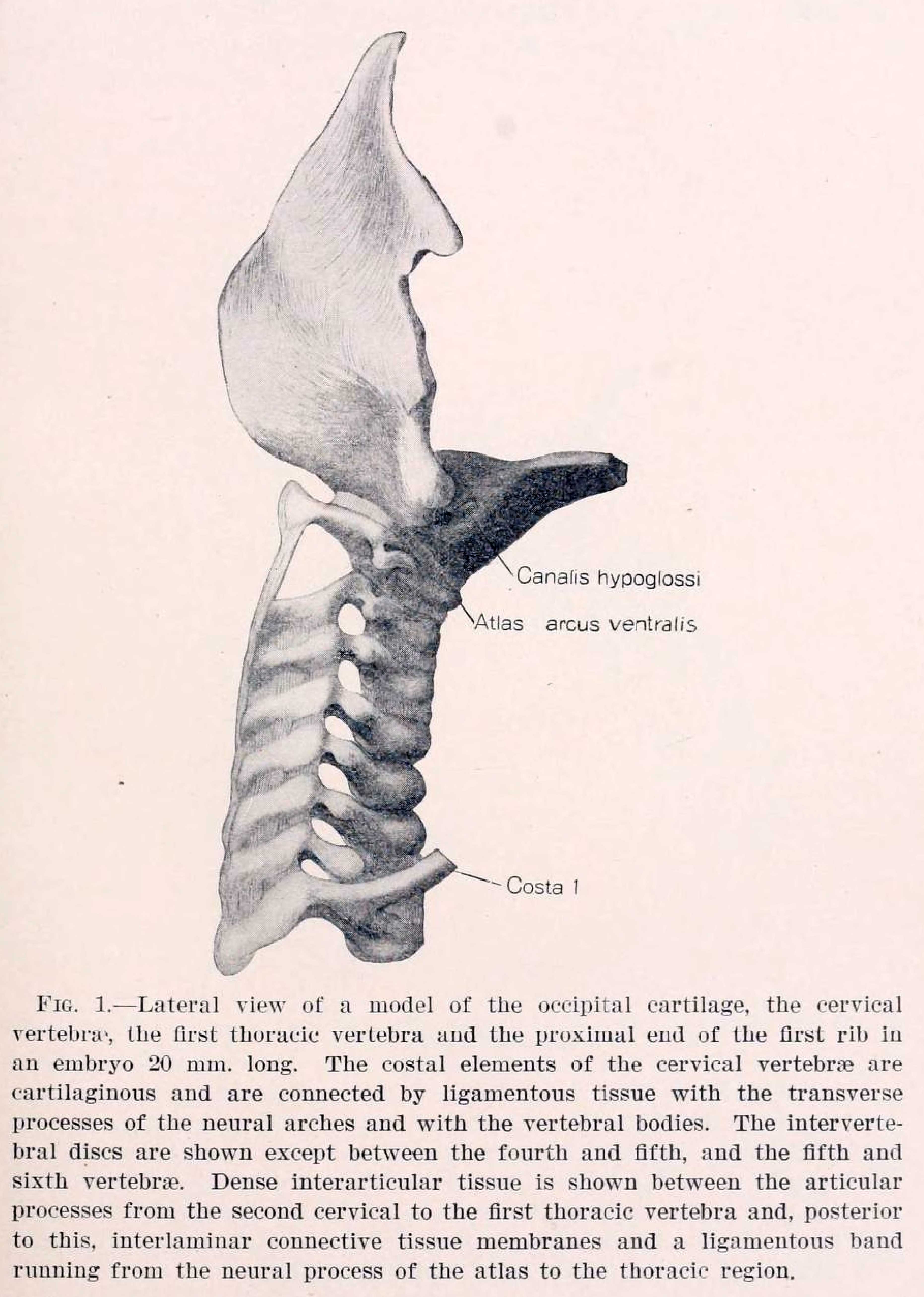
Fig. 1. Lateral view of a model of the occipital cartilage, the cervical vertebras the first thoracic vertebra aud the proximal end of the first rib in an embryo 20 mm. long. The costal elements of the cervical vertebrae are cartilaginous and are connected by ligamentous tissue with the transverse processes of the neural arches and with the vertebral bodies. The intervertebral discs are shown except between the fourth and fifth, and the fifth and sixth vertebnie. Dense interarticular tissue is shown between the articular processes from the second cervical to the first thoracic vertebra and, posterior to this, interlamiuar connectiye tissue membranes and a ligamentous band running from the neural process of the atlas to the thoracic region.
Reference
Bardeen CR. Early development of the cervical vertebrae and the base of the occipital bone in man. (1908) Amer. J Anat. 2: 182-186.
Cite this page: Hill, M.A. (2024, April 30) Embryology Bardeen1908a fig01.jpg. Retrieved from https://embryology.med.unsw.edu.au/embryology/index.php/File:Bardeen1908a_fig01.jpg
- © Dr Mark Hill 2024, UNSW Embryology ISBN: 978 0 7334 2609 4 - UNSW CRICOS Provider Code No. 00098G
File history
Click on a date/time to view the file as it appeared at that time.
| Date/Time | Thumbnail | Dimensions | User | Comment | |
|---|---|---|---|---|---|
| current | 16:17, 14 March 2018 | | 1,000 × 1,373 (76 KB) | Z8600021 (talk | contribs) | |
| 16:16, 14 March 2018 | Error creating thumbnail: File with dimensions greater than 12.5 MP | 3,725 × 5,227 (857 KB) | Z8600021 (talk | contribs) |
You cannot overwrite this file.
File usage
The following page uses this file: