File:Bardeen1905 plate13.jpg

Original file (1,000 × 1,337 pixels, file size: 85 KB, MIME type: image/jpeg)
Plate 13
Fig. 59. Section through the leg and foot of Embryo XVII 17, length 18 mm. The section does not pass through the cartilage of the 1st metatarsal.
Fig. 60. Section through the pubis, ischium, femur, fibula, calcaneus, cuboid and the 4th metatarsal cartilages of Embryo LXXIV Template:CE74, length 16 mm. 1}; diameters.
Figs. 61-65. Sections through the knee-joints of several embryos. 14 diam; 61, CCXXIX 229, length about 20 mm.; 62, LXXXVI 86, length 30 mm. ; 63, LXXV 75, length 30 mm.; 64 and 65, CXLV 145, length 33 mm.
Fig. 66. Longitudinal section .through the knee-joint, tibia. and foot-plate of Embryo CLXXV 175, length 13 mm.
Fig. 67. Section through the foot of Embryo CXLIV,144 length 14 mm.
Fig. 68. Section through the foot of Embryo LVII, length 23 mm. 62
Reference
Bardeen CR. Studies of the development of the human skeleton. (1905) Amer. J Anat. 4:265-302.
Cite this page: Hill, M.A. (2024, April 27) Embryology Bardeen1905 plate13.jpg. Retrieved from https://embryology.med.unsw.edu.au/embryology/index.php/File:Bardeen1905_plate13.jpg
- © Dr Mark Hill 2024, UNSW Embryology ISBN: 978 0 7334 2609 4 - UNSW CRICOS Provider Code No. 00098G
File history
Click on a date/time to view the file as it appeared at that time.
| Date/Time | Thumbnail | Dimensions | User | Comment | |
|---|---|---|---|---|---|
| current | 13:41, 10 September 2017 | | 1,000 × 1,337 (85 KB) | Z8600021 (talk | contribs) | |
| 13:41, 10 September 2017 | 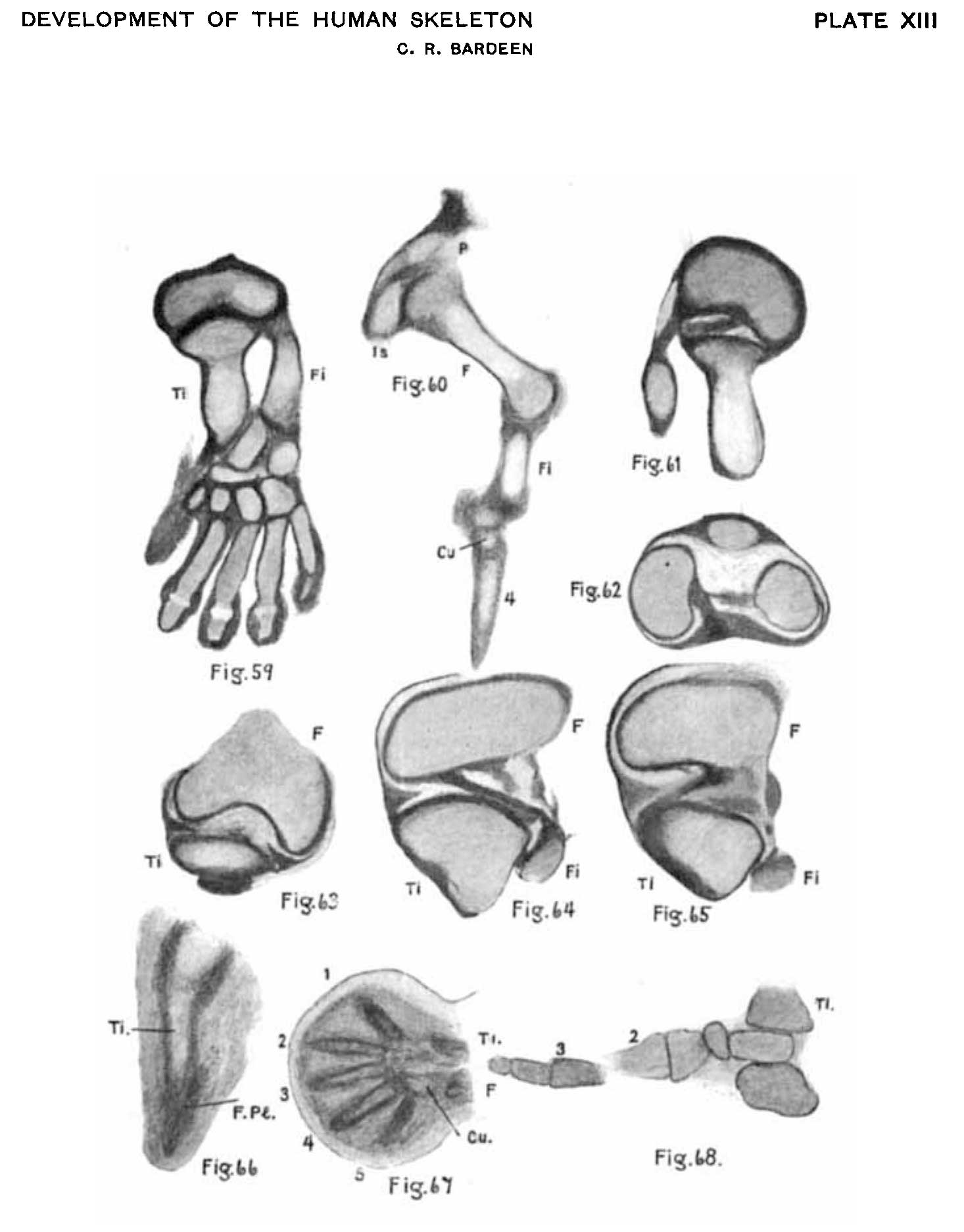 | 1,466 × 1,874 (109 KB) | Z8600021 (talk | contribs) | {{Ref-Bardeen1905}} |
You cannot overwrite this file.
File usage
The following page uses this file: