File:Bandler1983 fig07.jpg
From Embryology
Size of this preview: 704 × 600 pixels. Other resolution: 1,000 × 852 pixels.
Original file (1,000 × 852 pixels, file size: 223 KB, MIME type: image/jpeg)
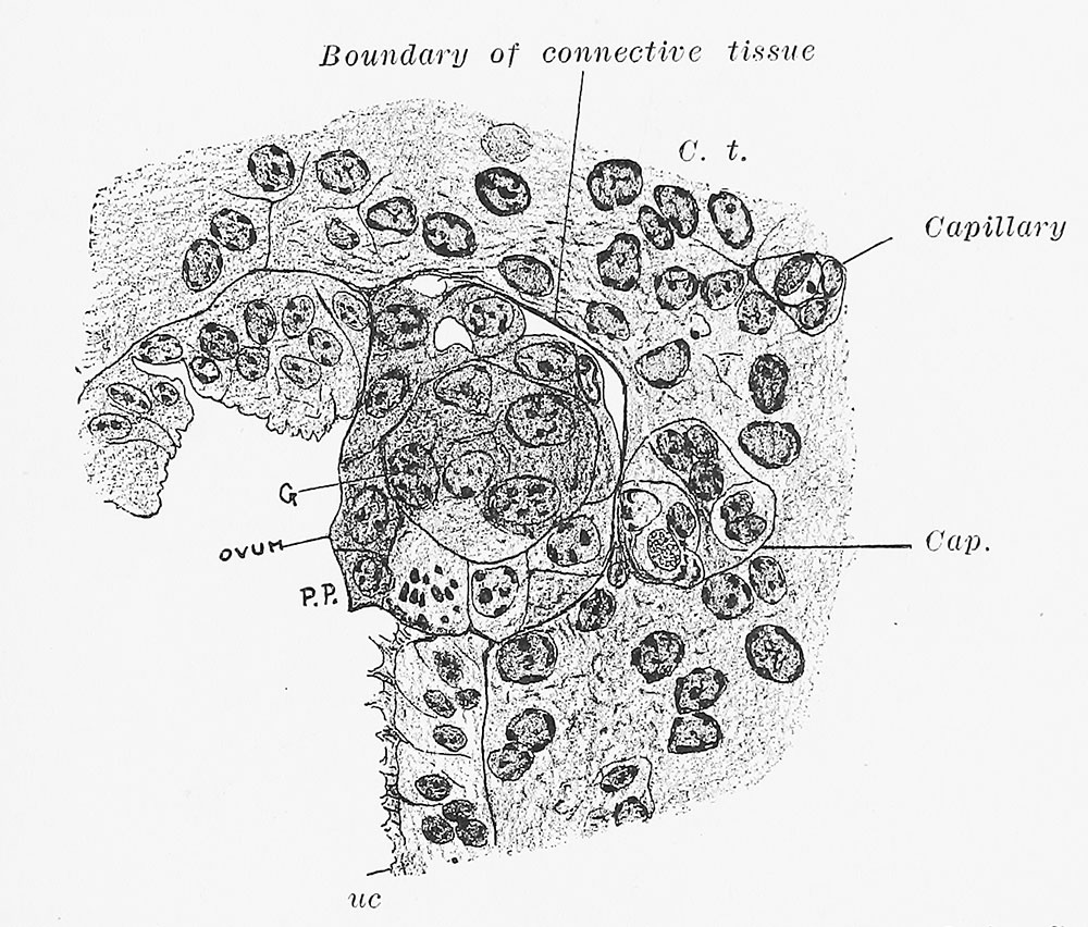
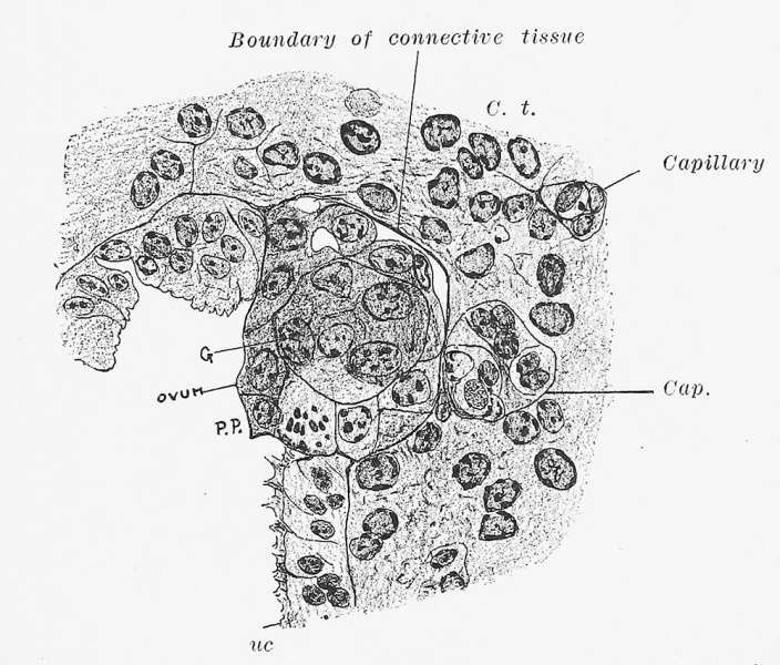
Fig. 7. A stage of embedding one-half hour later than Fig. 6
(v. Spee). more highly magnified. G, embryonal sphere. The ovum is surrounded by a symplasmatic zone.
Reference
Bandler SW. Uterine and Tubal Gestation (1893) William Wood & Company, New York.
Cite this page: Hill, M.A. (2024, May 7) Embryology Bandler1983 fig07.jpg. Retrieved from https://embryology.med.unsw.edu.au/embryology/index.php/File:Bandler1983_fig07.jpg
- © Dr Mark Hill 2024, UNSW Embryology ISBN: 978 0 7334 2609 4 - UNSW CRICOS Provider Code No. 00098G
File history
Click on a date/time to view the file as it appeared at that time.
| Date/Time | Thumbnail | Dimensions | User | Comment | |
|---|---|---|---|---|---|
| current | 23:11, 11 September 2018 | | 1,000 × 852 (223 KB) | Z8600021 (talk | contribs) | |
| 23:09, 11 September 2018 | 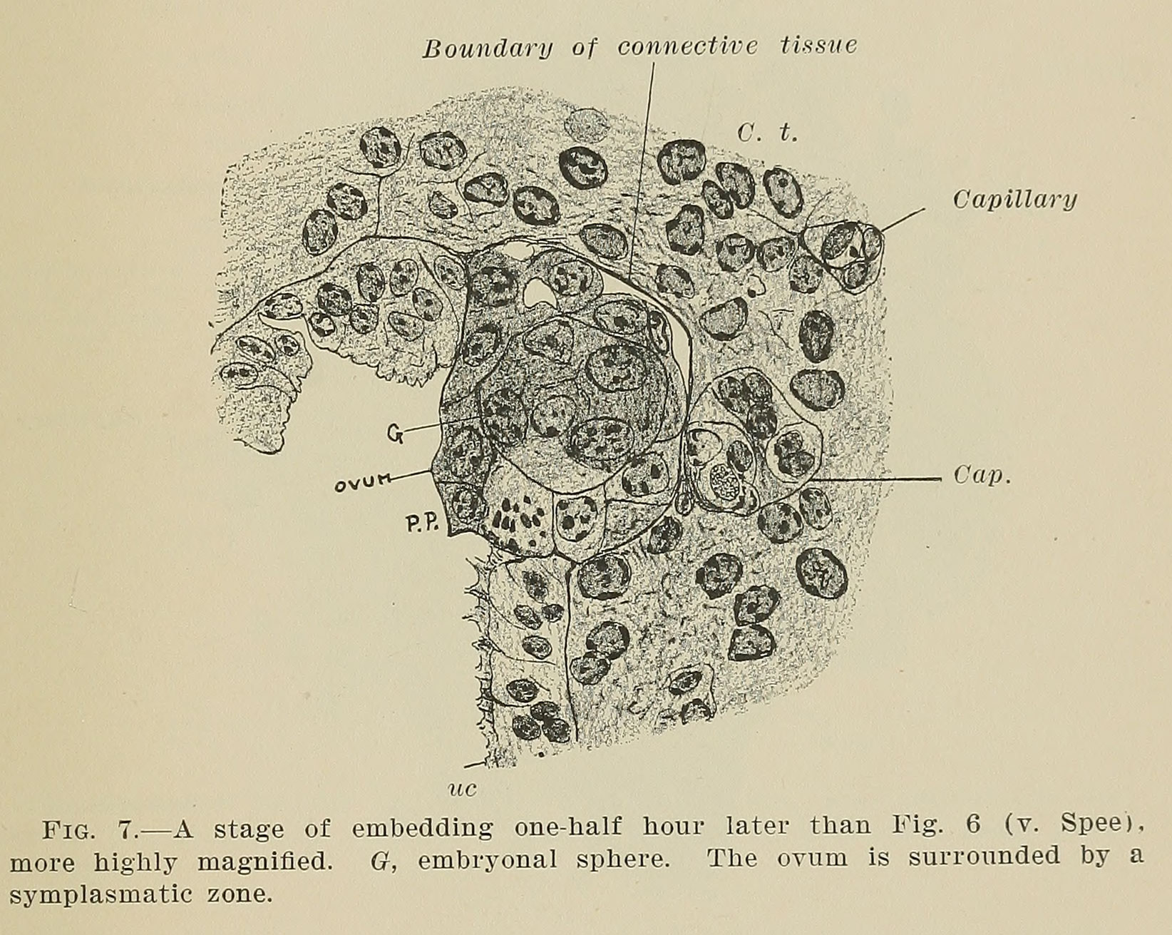 | 1,644 × 1,311 (441 KB) | Z8600021 (talk | contribs) | ===Reference=== {{Ref-Bandler1983}} {{Footer}} |
You cannot overwrite this file.
File usage
The following page uses this file: