File:Atwell1926 textfigA.jpg
Original file (869 × 700 pixels, file size: 191 KB, MIME type: image/jpeg)
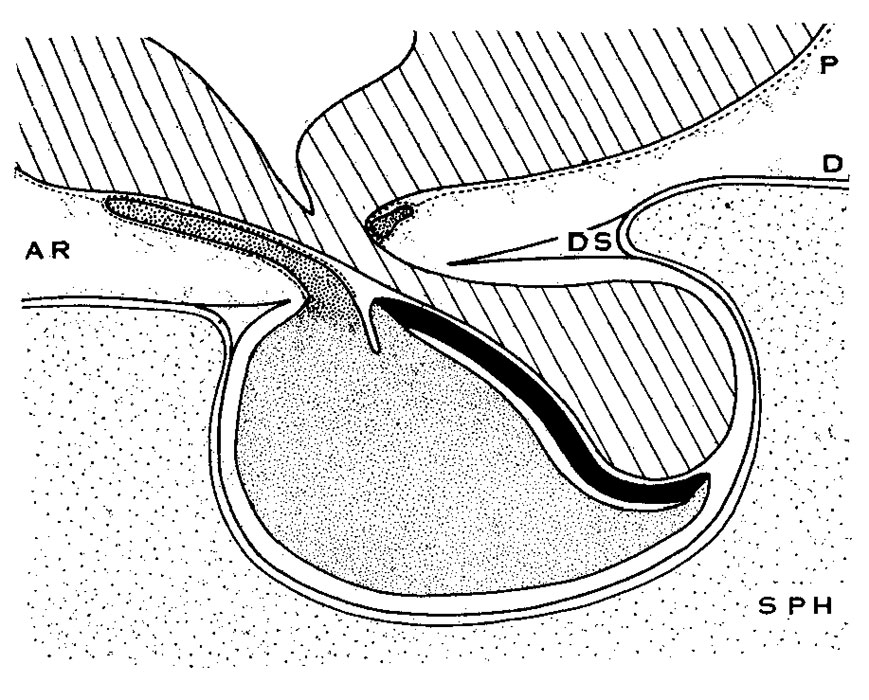
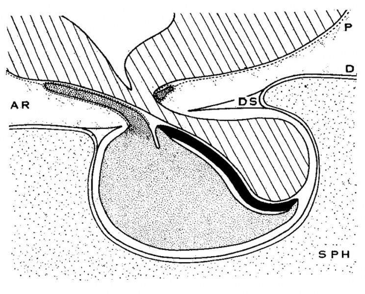
Text fig. A Diagrammatic sagittal section of hypophysis
Showing relation of pars tuberalis to the meninges. Lined, brain floor and pars neuralis; fine stipple, pars anterior proprior; coarse stipple, pars tnberalis; solid black, pars interniedia. SPH, sphenoid bone; P, pia lnator; D, (lura; DS, diaphragma sellae; AR, arachnoid spaces.
| Historic Disclaimer - information about historic embryology pages |
|---|
 |
Reference
Atwell WJ. The development of the hypophysis cerebri in man, with special reference to the pars tuberalis. (1926) Amer. J Anat. 37: 139-193.
Cite this page: Hill, M.A. (2024, May 19) Embryology Atwell1926 textfigA.jpg. Retrieved from https://embryology.med.unsw.edu.au/embryology/index.php/File:Atwell1926_textfigA.jpg
- © Dr Mark Hill 2024, UNSW Embryology ISBN: 978 0 7334 2609 4 - UNSW CRICOS Provider Code No. 00098G
File history
Click on a date/time to view the file as it appeared at that time.
| Date/Time | Thumbnail | Dimensions | User | Comment | |
|---|---|---|---|---|---|
| current | 16:59, 9 November 2016 | | 869 × 700 (191 KB) | Z8600021 (talk | contribs) | |
| 16:58, 9 November 2016 | 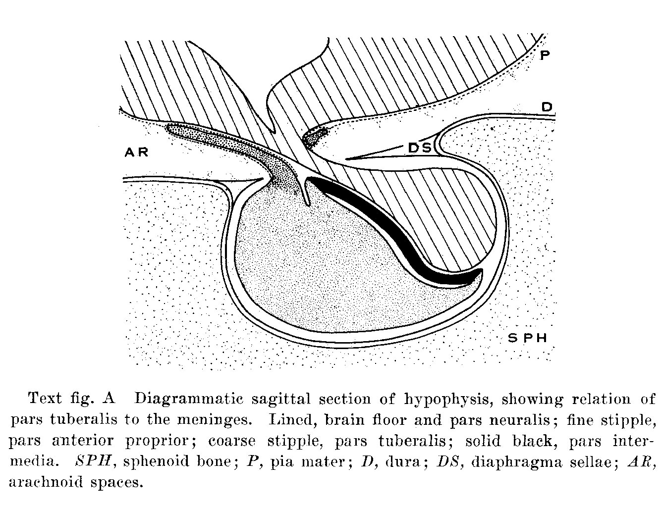 | 1,338 × 1,024 (308 KB) | Z8600021 (talk | contribs) | ==Text fig. A Diagrammatic sagittal section of hypophysis== Showing relation of pars tuberalis to the meninges. Lined, brain floor and pars neuralis; fine stipple, pars anterior proprior; coarse stipple, pars tnberalis; solid black, pars interniedia... |
You cannot overwrite this file.
File usage
The following page uses this file: