File:Atwell1926 plate06.jpg: Difference between revisions
mNo edit summary |
mNo edit summary |
||
| (One intermediate revision by the same user not shown) | |||
| Line 1: | Line 1: | ||
==Plate 6.== | ==Plate 6.== | ||
19 Wax—plalte reconstruction of the hypophysis from :1. 45-mm. human fetus (Huber collection, no. XVIII), viewed from above and in front. X 35. | |||
20 Reconstruction of the epithelial hypophysis from a 55»mm. human fetus (U.B.E.C., no. 25), viewed from above and in front. X 35. | |||
21 Reconstruction of epitllclial hypophysis from a 102-mm. human fetus (U.B.E.C., no. 17), viewed in rnidsagittal section; nasal, and at the right. X 35. | |||
Abbreviations (ml.l., an‘ro1'ior lobe p.int., pars intermedizi p.t., pars tuberalis rJ., residual lumen xx, incisure separating pars interme-di:1 and pars tuberalis 0.11.. ca,u<la,lly directed horn of pairs tuboralis i71f.n., infuiidibular 110t(‘.ll 11.1., neural lobe 1)., process of pars interinedizi g1'0win;_r up around neck of neural lobe 1:90 | |||
Latest revision as of 15:09, 9 November 2016
Plate 6.
19 Wax—plalte reconstruction of the hypophysis from :1. 45-mm. human fetus (Huber collection, no. XVIII), viewed from above and in front. X 35.
20 Reconstruction of the epithelial hypophysis from a 55»mm. human fetus (U.B.E.C., no. 25), viewed from above and in front. X 35.
21 Reconstruction of epitllclial hypophysis from a 102-mm. human fetus (U.B.E.C., no. 17), viewed in rnidsagittal section; nasal, and at the right. X 35.
Abbreviations (ml.l., an‘ro1'ior lobe p.int., pars intermedizi p.t., pars tuberalis rJ., residual lumen xx, incisure separating pars interme-di:1 and pars tuberalis 0.11.. ca,u<la,lly directed horn of pairs tuboralis i71f.n., infuiidibular 110t(‘.ll 11.1., neural lobe 1)., process of pars interinedizi g1'0win;_r up around neck of neural lobe 1:90
| Historic Disclaimer - information about historic embryology pages |
|---|
 |
Reference
Atwell WJ. The development of the hypophysis cerebri in man, with special reference to the pars tuberalis. (1926) Amer. J Anat. 37: 139-193.
Cite this page: Hill, M.A. (2024, May 18) Embryology Atwell1926 plate06.jpg. Retrieved from https://embryology.med.unsw.edu.au/embryology/index.php/File:Atwell1926_plate06.jpg
- © Dr Mark Hill 2024, UNSW Embryology ISBN: 978 0 7334 2609 4 - UNSW CRICOS Provider Code No. 00098G
File history
Click on a date/time to view the file as it appeared at that time.
| Date/Time | Thumbnail | Dimensions | User | Comment | |
|---|---|---|---|---|---|
| current | 15:07, 9 November 2016 | 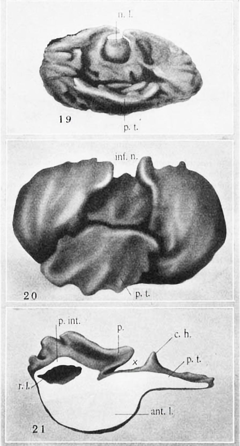 | 809 × 1,500 (136 KB) | Z8600021 (talk | contribs) | |
| 15:06, 9 November 2016 |  | 1,372 × 1,945 (215 KB) | Z8600021 (talk | contribs) |
You cannot overwrite this file.
File usage
The following page uses this file: