File:Atwell1926 plate04.jpg: Difference between revisions
mNo edit summary |
mNo edit summary |
||
| Line 2: | Line 2: | ||
12 Wax-plate reconstruction of epithelial hypophysis from a human embryo of 10.5 mm. (U.B.E.C., no.26), viewed from in front. X 65. | 12 Wax-plate reconstruction of epithelial hypophysis from a human embryo of 10.5 mm. (U.B.E.C., no.26), viewed from in front. X 65. | ||
13 | 13 Reconstruction of the epithelial hypophysis from a 14-mm human embryo (U.B.E.C., no. 46), viewed from in front. X 65. | ||
14 | 14 Reconstruction of the epithelial hypophysis from a 16-mm human embryo (Huber collection, 110. IV), viewed from in front. X 50. | ||
15 | 15 Reconstruction of the epithelial hypophysis from a 17-mm human embryo (U.B.E.C., no. 42), viewed from in front. X 65. | ||
Abbreviations a.c., ‘anterior chamber’ l.l., lateral lobe | Abbreviations a.c., ‘anterior chamber’ l.l., lateral lobe; d, diverticulum st, epithelial stalk inf. n. infundibular notch | ||
{{Historic Disclaimer}} | {{Historic Disclaimer}} | ||
Latest revision as of 08:51, 15 November 2016
Plate 4
12 Wax-plate reconstruction of epithelial hypophysis from a human embryo of 10.5 mm. (U.B.E.C., no.26), viewed from in front. X 65.
13 Reconstruction of the epithelial hypophysis from a 14-mm human embryo (U.B.E.C., no. 46), viewed from in front. X 65.
14 Reconstruction of the epithelial hypophysis from a 16-mm human embryo (Huber collection, 110. IV), viewed from in front. X 50.
15 Reconstruction of the epithelial hypophysis from a 17-mm human embryo (U.B.E.C., no. 42), viewed from in front. X 65.
Abbreviations a.c., ‘anterior chamber’ l.l., lateral lobe; d, diverticulum st, epithelial stalk inf. n. infundibular notch
| Historic Disclaimer - information about historic embryology pages |
|---|
 |
Reference
Atwell WJ. The development of the hypophysis cerebri in man, with special reference to the pars tuberalis. (1926) Amer. J Anat. 37: 139-193.
Cite this page: Hill, M.A. (2024, May 13) Embryology Atwell1926 plate04.jpg. Retrieved from https://embryology.med.unsw.edu.au/embryology/index.php/File:Atwell1926_plate04.jpg
- © Dr Mark Hill 2024, UNSW Embryology ISBN: 978 0 7334 2609 4 - UNSW CRICOS Provider Code No. 00098G
File history
Click on a date/time to view the file as it appeared at that time.
| Date/Time | Thumbnail | Dimensions | User | Comment | |
|---|---|---|---|---|---|
| current | 16:34, 9 November 2016 |  | 1,200 × 1,177 (152 KB) | Z8600021 (talk | contribs) | |
| 16:34, 9 November 2016 | 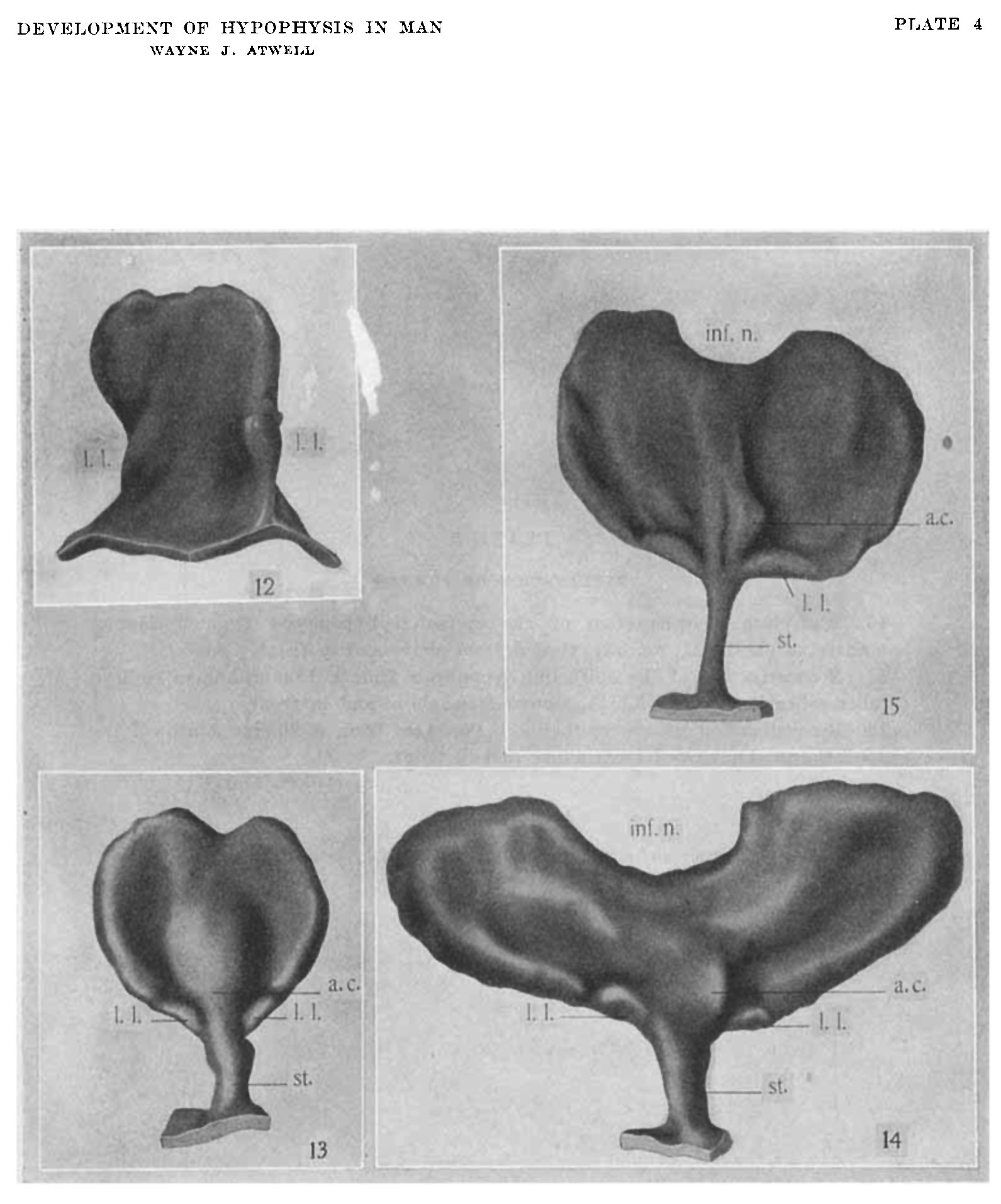 | 1,591 × 1,913 (242 KB) | Z8600021 (talk | contribs) |
You cannot overwrite this file.
File usage
The following page uses this file: