File:Atwell1918 fig34.jpg: Difference between revisions
No edit summary |
mNo edit summary |
||
| (2 intermediate revisions by the same user not shown) | |||
| Line 1: | Line 1: | ||
==Fig. 34 Model of hypophysis from twenty-eight-day embryo=== | |||
X 25. Brain wall has been removed so as to present a dorsal view of the gland. The two nasal horns and the two caudal horns of pars tuberalis have fused. p.t., pars tubercles; 2)., process growing up to surround neck of neural lobe; n.I., neural lobe | |||
{{Historic Disclaimer}} | |||
See also {{Ref-Atwell1926}} | |||
'''Links:''' [[Endocrine - Pituitary Development|Pituitary Development]] | [[Rabbit Development]] | |||
===Reference=== | |||
{{Ref-Atwell1918}} | |||
{{Footer}} | |||
[[Category:Pituitary]][[Category:Rabbit]][[Category:1910's]] | |||
Latest revision as of 11:12, 16 November 2016
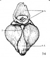
Fig. 34 Model of hypophysis from twenty-eight-day embryo=
X 25. Brain wall has been removed so as to present a dorsal view of the gland. The two nasal horns and the two caudal horns of pars tuberalis have fused. p.t., pars tubercles; 2)., process growing up to surround neck of neural lobe; n.I., neural lobe
| Historic Disclaimer - information about historic embryology pages |
|---|
| Pages where the terms "Historic" (textbooks, papers, people, recommendations) appear on this site, and sections within pages where this disclaimer appears, indicate that the content and scientific understanding are specific to the time of publication. This means that while some scientific descriptions are still accurate, the terminology and interpretation of the developmental mechanisms reflect the understanding at the time of original publication and those of the preceding periods, these terms, interpretations and recommendations may not reflect our current scientific understanding. (More? Embryology History | Historic Embryology Papers) |
See also Atwell WJ. The development of the hypophysis cerebri in man, with special reference to the pars tuberalis. (1926) Amer. J Anat. 37: 139-193.
Links: Pituitary Development | Rabbit Development
Reference
Atwell WJ. The development of the hypophysis cerebri of the rabbit (Lepus Cuniculus L.). (1918) Amer. J Anat. 24(2): 271-337
Cite this page: Hill, M.A. (2024, May 15) Embryology Atwell1918 fig34.jpg. Retrieved from https://embryology.med.unsw.edu.au/embryology/index.php/File:Atwell1918_fig34.jpg
- © Dr Mark Hill 2024, UNSW Embryology ISBN: 978 0 7334 2609 4 - UNSW CRICOS Provider Code No. 00098G
File history
Click on a date/time to view the file as it appeared at that time.
| Date/Time | Thumbnail | Dimensions | User | Comment | |
|---|---|---|---|---|---|
| current | 11:08, 16 November 2016 |  | 353 × 406 (32 KB) | Z8600021 (talk | contribs) | |
| 11:08, 16 November 2016 |  | 1,000 × 1,351 (357 KB) | Z8600021 (talk | contribs) |
You cannot overwrite this file.
File usage
There are no pages that use this file.
